
Innovation ingredients
Skin care
KEYWORDS
anti-aging;
bioavailability;
well-being
Peer Reviewed
Mimicking Bioelectrical Stimulation: Enhancing Intracellular Uptake and Skin Vitality via Plant-Based Glycoconjugates
Ana Gallego, Victoria Vidal, Sandra Ruiz, Pau Riera, Daniel Luna, Xinwei Zhao, Maria Mas, Marta Gibert, Laura Cano, Sara Laplana, Tarik Ruiz, Laura Facchini, Alejandro Guirado, Òscar Expósito*
*Corresponding author
Vytrus Biotech, Terrassa, Spain
ABSTRACT: Efficient and non-invasive active ingredient delivery remains a major challenge in cosmetic skin treatments. Here, we studied a bioinspired self-delivery mechanism derived from Hylocereus undatus stem cells (Hu) that mimics electroporation through biologically driven membrane permeabilization.
In vitro, Hu promoted sustained intracellular dye uptake while preserving cell viability, increased extracellular ATP (eATP) release and stimulated VEGF secretion. Mechanistically, these effects appear to involve eATP-mediated P2X receptor activation via Hu glycoconjugates, enhancing eATP signaling and facilitating the cellular entry of bioactive compounds. In vivo studies confirmed efficacy comparable to needle-free mesotherapy in the eye contour area and significant improvements in key skin-aging parameters.
Introduction
Introduction
The skin is a sophisticated barrier that shields against environmental aggressors while preserving internal balance. Beyond its classical protective role the skin is increasingly recognized as a bioelectrical tissue capable of generating and responding to endogenous and exogenous electric signals. These bioelectrical phenomena regulate key processes such as wound healing, inflammation, and tissue regeneration, and are emerging as novel mechanisms for cosmetic delivery (1-4). Notably, ATP-mediated signaling through P2X receptors has been implicated in the propagation of bioelectrical cues and the regulation of these processes (1,5).
Electroporation, or needle-free mesotherapy, uses short, high-voltage electrical pulses to transiently disrupt cell membranes and facilitate the uptake of bioactive molecules through the skin (6-9). Beyond delivery, electroporation and other electrostimulation-based therapies have demonstrated regenerative and rejuvenating benefits, promoting keratinocyte and fibroblast proliferation, growth factor secretion, collagen synthesis, ATP production and cellular migration while modulating inflammatory responses (3,10-13). Despite these benefits, electroporation remains limited by device complexity, safety concerns, and the lack of standardized guidelines (8,11,14-17).
Here, we describe a bioinspired self-delivery system derived from Hylocereus undatus stem cells (Hu) that safely mimics electroporation through biologically driven membrane permeabilization and without the need for external devices. We hypothesized that the unique glycoconjugate-rich composition of Hu potentiates ATP-dependent P2X signalling, promoting transient membrane permeabilization and intracellular delivery of bioactive compounds.
Materials and Methods
In vitro studies
Hylocereus undatus cell cultures
Totipotent cell cultures of Hylocereus undatus (Hu)were established in modified MS medium and incubated in darkness under continuous agitation to promote biomass accumulation and enhance the production of polyphenols and glycoconjugates.
Experimental system and culture conditions
HaCaT (CLS, Germany) were maintained in complete culture medium (DMEM + 10% FBS) and HDF (Innoprot, Spain) were cultured in fibroblast medium (2% FBS). Cells were seeded at 105 cells/well in 96-well plates and cultured under standard conditions (37ºC, 5% CO2).
HaCaT cell electroporation: protocol development
Electroporation was performed using a Gene Pulser Xcell system (Bio-Rad, USA). HaCaT cells were treated with 1 µM BLOCK-iT™ dye and exposed to voltages ranging from 80-160V and pulse durations of 15-25 ms to establish optimal conditions. Cell viability and dye penetration were analysed using a CytoFLEX S Flow Cytometer (Beckman Coulter, USA). Results were expressed as a % of variation relative to C- (non-electroporated dye-treated cells).
Electroporation-mimetic mechanism assessment
HaCaT cells were treated with Hu (1, 5, 20% (v/v)), ATP (5, 10 mM), or electroporated under optimal conditions (110 V, 20 ms). Membrane permeability (evaluated by 1 µM BLOCK-iT™ dye permeabilization analysed by flow cytometry (n=6) and quantified as median fluorescence intensity (MFI)), cell viability (MTT assay), and VEGF production (n=4) (Human VEGF DuoSet ELISA Kit, R&D Systems, USA) were assessed at different incubation times: 1, 4, or 24 hours. Dye permeabilization data were represented as median fluorescence intensity (MFI), cell viability was expressed as percentage variation relative to the C− (non-electroporated, dye-treated cells), and VEGF values were normalized to cell viability. Dye incorporation was visualized using a high-content imaging system (Operetta CLS, PerkinElmer, MA, USA), with nuclei counterstained with 2 μM Hoechst.
Measurement of eATP in HaCaT and HDF Cells
HDF and HaCaT cells were treated with Hu (0.3, 1.25, 5% (v/v)) and incubated for 24 h. Then, extracellular ATP (eATP) levels were measured using the RealTime-Glo™ Extracellular ATP Assay Kit (Promega, USA) (n=3). The results were expressed as a % of variation relative to C- (untreated cells).
Statistical analysis
Statistical significance was determined using a two-tailed Student’s t-test, while variance homogeneity was evaluated using Fisher’s F-test. A p-value < 0.05 was considered indicative of statistical significance.
In vivo studies
Comparative evaluation with a needle-free mesotherapy device
A double-blind comparative study was conducted to evaluate the clinical effects of Hu versus a needle-free mesotherapy device for at-home use combining electroporation, electrical muscle stimulation (EMS), radiofrequency (RF), and LED photon therapy (415nm blue light, 90kHz, 10W). Forty-four female and male subjects (45-70 years) with mild-to-moderate signs of aging (crow’s feet wrinkles, fine lines, eyelid sagging and under eye-bags) were randomized into two hemi-face treatment groups (n=22): 3% Hu formulation versus placebo, and device plus placebo versus device plus conductive cream containing hyaluronic acid, vitamin C, and collagen (hereafter, device cream). The device-treated group applied treatments to the eye contour area four times weekly (10 minutes/eye) as per the provider’s instructions, whereas the formulation-only group applied treatments twice daily with a 3–5-minute massage per eye. Assessments were performed at baseline (T0), day 21 (T21) and day 42 (T42), and included eye-contour profilometry (PRIMOS 3D, Canfield Scientific, Netherlands), eyelid sagging analysis from standardized VISIA®-CR images (Canfield Scientific), and eye-opening angle measurements using Visioface®1000D (Courage+Khazaka, Germany).
Short- and long-term effects on skin aging parameters
Two additional double-blind clinical studies were conducted to evaluate the short- and long-term effects of Hu formulations on skin aging parameters. In the first study, forty female subjects (40–65 years) with dry skin tendency and mild-to-moderate signs of facial aging were randomized into two groups (n=20) and received a 2% Hu formulation or a placebo. Formulations were applied twice daily over the entire face and assessments, performed at T0, day 14 (T14) and day 28 (T28), included skin radiance (CM-700D, Konica-Minolta, Japan) and moisturization (Corneometer®, Courage+Khazaka). In the second study, 60 female and male subjects (40–65 years) with facial skin sagging and mild-to-moderate signs of facial aging were randomized into two groups (n=30) and received either 1.5% Hu formulations or placebos. Treatments were applied twice daily over the entire face and measurements were performed at T0, T28 and day 56 (T56). Skin elasticity (R2), firmness (R0) and fatigue (R9) were measured via Cutometer® MPA580 (Courage+Khazaka).
Statistical analysis
Instrumental data were analysed via two-way Student’s t-test for paired data. A p-value < 0.05 was considered indicative of statistical significance.
Results and Discussion
In vitro studies
HaCaT cell electroporation: protocol development
The electroporation conditions for HaCaT cells were optimized to maximize BLOCK-iT™ dye uptake while preserving cell viability. As shown in Figure 1, electroporation efficiency increased with voltage and pulse duration, reaching a maximum at 130 V. However, these conditions significantly reduced cell viability, highlighting the inverse relationship between electroporation efficiency and cell survival.
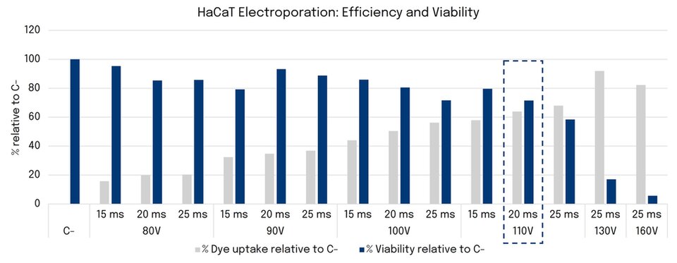
Figure 1. Dye uptake (%) and viability (%) of HaCaT cells following electroporation optimization between 80–160 V and 15–25 ms. C−: non-electroporated dye-treated cells with 1 µM BLOCK-iT™.
Based on these results, 110 V/20 ms was selected as the optimal condition, providing a balance between electroporation efficiency (64%) and cell viability (72%). These findings are consistent with previous reports in HaCaT cells showing near 80% viability following electroporation at 175 V/10 ms (6,18).
Electroporation-mimetic mechanism assessment
Dye uptake as a measure of membrane permeabilization
As shown in Figures 2 and 3, both Hu and eATP promoted intracellular BLOCK-iT™ uptake in HaCaT cells. Hu induced a concentration- and time-dependent increase in dye uptake, reaching a maximum MFI of 26,419 at 20% (v/v) after 24 h (+356% vs 1 h). Dye uptake was also observed at 1% and 5% Hu. In addition, eATP enhanced dye uptake, with the highest response observed at 10 mM after 4h and a sustained increase at 5 mM over time, reaching an MFI of 19,061 after 24 h. Despite these effects, the highest dye uptake was achieved by electroporation (110 V/20 ms), which yielded an MFI of 83,262, corresponding to a 3.15-fold and 2.92-fold higher signal than the maximum responses induced by Hu and eATP, respectively.
Unlike electroporation, in which membrane pores begin to close within seconds and fully reseal after 10 minutes (19), Hu treatment may involve prolonged pore opening or repeated cycles of opening and closing, allowing a cumulative intracellular entry throughout the incubation period (20,21).
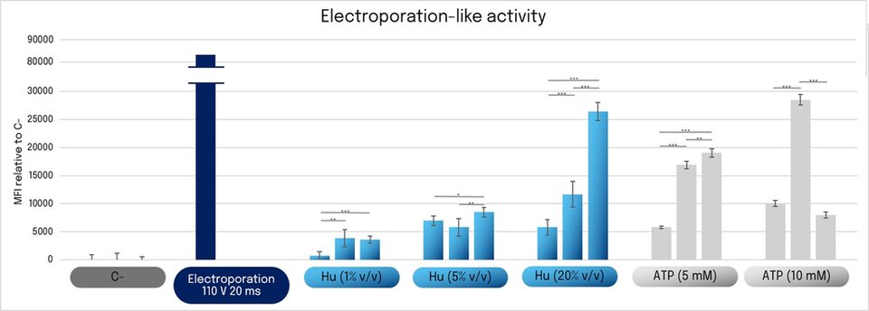
Figure 2. Electroporation-like activity in HaCaT. Median fluorescence intensity (MFI) of internalized BLOCK-iT™ after electroporation (110V/20ms) or following incubation with Hu (1, 5, 20% (v/v)) or ATP (5,10 mM) for 1, 4, and 24 h. Data represent the mean ± SD of n = 6. Asterisks indicate statistically significant differences (*p < 0.05, **p < 0.01, ***p < 0.001).
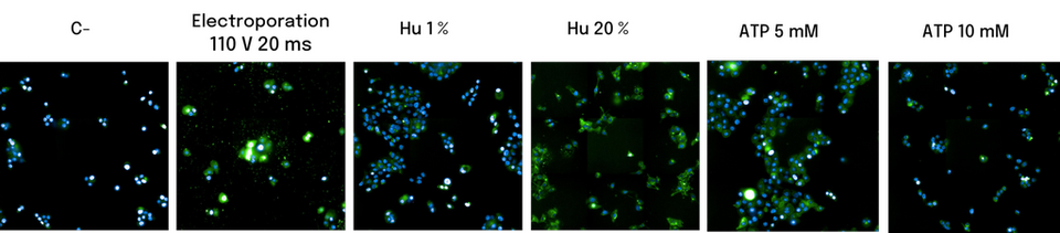
Figure 3. Electroporation-like activity in HaCaT after 24 h of Hu (1, 20% (v/v)) or ATP (5,10 mM) incubation (high-content imaging) compared to electroporation (110 V/20 ms). Green: BLOCK-iT™ uptake; blue: Hoechst nuclear staining.
As previously mentioned, the evaluation of electroporation-like activity requires simultaneous assessment of membrane permeabilization and cell viability. Therefore, cell viability was measured at 1, 4, and 24 h after electroporation or following 1-, 4-, and 24-h incubations with Hu or eATP (Figure 4).
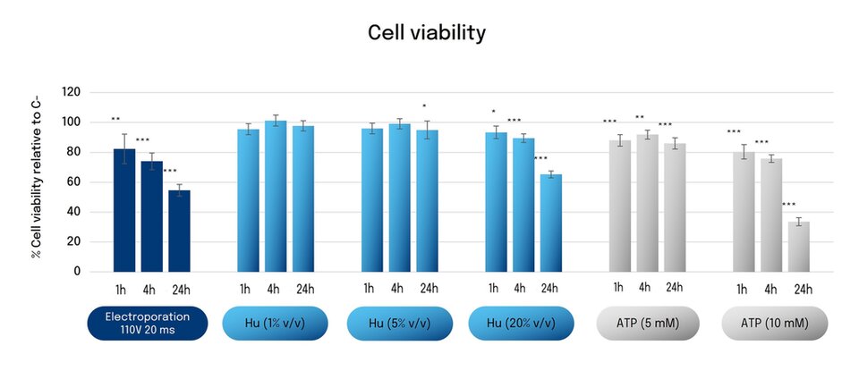
Figure 4. Cell viability (% relative to C−) at 1, 4, and 24 h following electroporation (110 V/20 ms), Hu (1–20%), or eATP (5–10 mM). Asterisks indicate statistically significant differences vs C− (*p < 0.05, **p < 0.01, ***p < 0.001). Data represent the mean ± SD of n= 6.
Electroporation moderately reduced cell viability after 1–4 h, followed by a marked decline to 55% at 24 h, suggesting delayed cell damage associated with membrane permeabilization (22-24). Hu at 1% and 5% did not significantly affect cell viability, whereas 20% Hu reduced viability after 24 h.Similarly, 5 mM eATP had no effects, while 10 mM reduced viability to 34% after 24 h. Unlike electroporation, eATP probably induces membrane permeabilization through P2XR activation and pore formation (5,20,21, 25-29), while the cytotoxicity observed at 10 mM is likely due to supraphysiological ATP exposure (30).
VEGF induction as an electroporation-mimetic response
VEGF production was evaluated as a marker of cellular stress and regenerative responses (31). As shown in Figure 5, VEGF secretion increased over time in control cells, reaching 179 pg/mL at 24 h. Electroporation initially reduced VEGF levels but increased secretion by 29% after 24 h. In contrast, 5 mM eATP induced a 69% increase in VEGF production at 4 h, although this effect was not maintained at 24 h. Hu induced a dose- and time-dependent increase in VEGF secretion, reaching 58% and 187% above control levels at 4 and 24 h, respectively. P2X7R-mediated activation and VEGF release was reported in various cell types (32).
Overall, VEGF secretion was not directly proportional to membrane permeabilization, indicating the involvement of additional mechanisms, probably related with Hu-derived compound uptake.
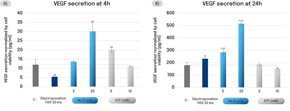
Figure 5. VEGF secretion in HaCaT at (A) 4 h and (B) 24 h after electroporation (110 V, 20 ms), Hu (5, 20%) or ATP (5, 10 mM) treatments. Data is normalized by cell viability and expressed as pg/mL; bars represent the mean ± SD of n=4. Asterisks indicate statistically significant differences vs C- (*p < 0.05, **p < 0.01, ***p < 0.001).
eATP release as an indicator of P2X receptor activation
To further assess the potential involvement of P2X pathway activation by Hu, eATP release was assessed in HDF and HaCaT cells.
As shown in Figure 6, Hu induced a dose-dependent increase in eATP release in both cell lines, with a stronger response in HaCaT cells (up to 116%) than in HDF cells (up to 42%), consistent with the higher expression of P2X receptors reported in keratinocytes (33).
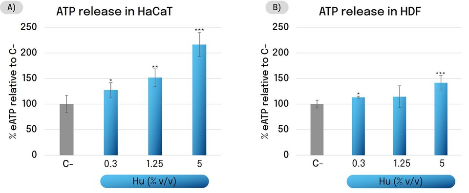
Figure 6. eATP release in (A) HaCaT and (B) HDF cells after 24 h Hu treatment (0.3–5%). Results expressed as % vs. untreated control (C-). Asterisks indicate statistically significant differences compared to C- (*p < 0.05, **p < 0.01, ***p < 0.001). Data represent the mean ± SD of n = 3.
To better understand the proposed mechanism of action, it is important to consider the unique composition of Hu. Produced through a biotechnological process, Hu is enriched in glycoconjugates (~80% of its protein content), together with complex sugars, polyphenols, and pigments (34). We hypothesize that the combined activity of these bioactive components underlies the electroporation-mimetic effects observed in this study. In particular, glycoconjugates have been reported to enhance P2X receptor signalling through positive allosteric modulation (35–39). Together with the observed increases in eATP release, membrane permeabilization, and VEGF production, these findings support a model in which Hu potentiates P2X activation and promotes transient membrane permeabilization and pore formation (5,32). This may facilitate the intracellular delivery of Hu-derived bioactive compounds, consistent with the reported use of P2X receptors as a gateway for intracellular delivery (21,40). Increased intracellular availability of these compounds could contribute to the regenerative activity associated with Hu.
In vivo studies
Comparative evaluation with a needle-free mesotherapy device
The electroporation-like effect of Hu was evaluated in vivo by comparing Hu 3% with three control conditions: placebo, placebo combined with a needle-free mesotherapy device, and a device-specific cream delivered using the same device.
Hu 3% induced significant cumulative improvements at all evaluated periocular parameters after 21 and 42 days (Figure 7). After 42 days of treatment, wrinkle depth decreased by 13.0%, eye bag volume by 8.9%, and eyelid sagging by 0.21 mm, while the eye-opening angle increased by 4.8%. Notably, Hu 3% outperformed the placebo and the device plus placebo across all parameters, with no significant differences versus the device combined with its active cream. The superior efficacy of the device plus placebo versus placebo alone confirms the contribution of electrostimulation to skin improvement, while the addition of the active cream further enhanced efficacy, likely by improving current conduction and active delivery. Collectively, these findings suggest that Hu can reproduce the benefits of a complete mesotherapy treatment despite being applied without any delivery device, supporting the self-delivery and biological activity of its bioactive compounds.
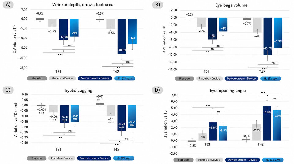
Figure 7. Comparative evaluation of Hu 3%’s and a needle-free mesotherapy device’s effects on age-related eye contour parameters: (A) wrinkle depth in the crow’s feet area, (B) eye bag volume, (C) eyelid sagging, and (D) eye-opening angle after 21 and 42 days of treatment. Asterisks indicate statistically significant differences (*p < 0.05, **p < 0.01, ***p < 0.001).
Short- and long-term effects on skin aging parameters
Long-term treatment with Hu at 1.5% significantly improved skin firmness, enhanced skin elasticity, and reduced skin fatigue (Figure 9). These effects became more pronounced over time, supporting the ability of Hu to improve both the appearance and biomechanical properties of the skin through sustained use.
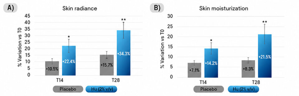
Figure 8. Short-term effects of Hu on (A) skin radiance and (B) moisturization. Graphs show the mean percentage ± SE change from baseline (T0) after 14 and 28 days of treatment. Asterisks indicate statistically significant differences compared to the placebo group (*p < 0.05, **p < 0.01).
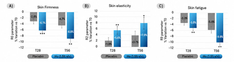
Figure 9. Long-term effects of Hu on (A) skin firmness, (B) skin elasticity, and (C) skin fatigue. Graphs show the mean percentage ± SE change from baseline (T0) after 28 and 56 days of treatment. Asterisks indicate statistically significant differences compared to the placebo (*p < 0.05, **p < 0.01, ***p < 0.001).
Conclusion
Altogether, these results position Hylocereus undatus stem cells as a novel glycoconjugate-rich self-delivery system that promotes an electroporation-mimetic response through enhanced transient membrane permeabilization. This mechanism may facilitate the intracellular delivery of Hu-derived bioactive compounds while preserving cell viability, leading to the activation of regenerative pathways in vitro and significant improvements in multiple signs of skin aging in vivo. Notably, Hu achieved efficacy comparable to needle-free mesotherapy in the eye contour area, highlighting its potential as a non-invasive strategy for skin rejuvenation.
Ethics statement: All clinical studies were conducted in accordance with the Declaration of Helsinki, and written informed consent was obtained from all participants.
Conflict of Interest Statement: The authors are the developers and intellectual property holders of the active ingredient evaluated in this study. However, all in vitro and in vivo studies were conducted by independent third-party laboratories. The authors were not involved in the execution of the experimental procedures. This potential conflict of interest is disclosed in the interest of transparency.
About the Author
Bachelor’s Degree in Biology and PhD in Plant Biotechnology from the Faculty of Pharmacy of the University of Barcelona (Spain). There he met Albert Jané and founded Vytrus Biotech in 2009 to develop active ingredients for skin and hair care through plant biotechnology. His passion for plants has led him to be the author of more than 14 scientific articles and 7 patents in the cosmetic and pharmaceutical sector. He has participated as a speaker in +35 international academic conferences, as a professor in 7 Masters and specialization courses in the fields of biotechnology and entrepreneurship and is a member of prestigious associations such as SEQC, SCS and IFSCC. With Vytrus, he has won +30 international awards in innovation & sustainability in the cosmetic sector.

Òscar Expósito*
*Corresponding author
Vytrus Biotech, Terrassa, Spain

References and notes
- Yu, S. M., Granick, S., Electric spiking activity in epithelial cells, Proceedings of the National Academy of Sciences of the United States of America, 122 (2025) e2427123122, https://doi.org/10.1073/pnas.2427123122
- McLaughlin, K. A., Levin, M., Bioelectric signaling in regeneration: Mechanisms of ionic controls of growth and form, In Developmental Biology, Elsevier Inc., 433 (2018) 177–189, https://doi.org/10.1016/j.ydbio.2017.08.032
- Poltawski, L., Watson, T., Bioelectricity and microcurrent therapy for tissue healing–a narrative review, In Physical Therapy Reviews, Taylor and Francis Ltd., 14 (2009) 104–114, https://doi.org/10.1179/174328809X405973
- Flores-Muñoz et al., Restraint of human skin fibroblast motility, migration, and cell surface actin dynamics, by pannexin 1 and p2x7 receptor signaling, International Journal of Molecular Sci-ences, 22 (2021) 1–18, https://doi.org/10.3390/ijms22031069
- Zimmermann, H., Extracellular ATP and other nucleotides—ubiquitous triggers of intercellular messenger release, In Purinergic Signalling, Springer Netherlands, 12 (2016) 25–57, https://doi.org/10.1007/s11302-015-9483-2
- Skorupska, S., Grabowska-Jadach, I., Cytotoxicity studies of quantum dots with the electro-poration method, Bioelectrochemistry, 126 (2019) 86–91, https://doi.org/10.1016/j.bioelechem.2018.11.011
- Shah et al., Innovative transdermal drug delivery systems: Benefits, challenges, and emerging applications, In BMEMat. John Wiley and Sons Inc., 2025, Article e70001, https://doi.org/10.1002/bmm2.70001
- Dermol-Černe, J., Pirc, E., Miklavčič, D., Mechanistic view of skin electroporation–models and dosimetry for successful applications: an expert review, In Expert Opinion on Drug Delivery, Taylor and Francis Ltd., 17 (2020) 689–704, https://doi.org/10.1080/17425247.2020.1745772
- Zorec, B., Becker, S., Reberšek, M., Miklavčič, D., & Pavšelj, N., Skin electroporation for transdermal drug delivery: The influence of the order of different square wave electric pulses, International Journal of Pharmaceutics, 457 (2013) 214–223, https://doi.org/10.1016/j.ijpharm.2013.09.020
- Urabe et al., Effects of pulsed electrical stimulation on growth factor gene expression and proliferation in human dermal fibroblasts, Molecular and Cellular Biochemistry, 476 (2021) 361–368. https://doi.org/10.1007/s11010-020-03912-6
- Xu, X., Zhang, H., Yan, Y., Wang, J., Guo, L., Effects of electrical stimulation on skin surface, In Acta Mechanica Sinica/Lixue Xuebao, Springer Verlag, 37 (2021) 1843–1871, https://doi.org/10.1007/s10409-020-01026-2
- Golberg et al., Skin rejuvenation with non-invasive pulsed electric fields, Scientific Reports, 5 (2015) 10187, https://doi.org/10.1038/srep10187
- Tripathi, S., Diverse Applications of Electrostimulation and its Future Prospects, Journal of Biomedical Sciences, 11 (2022) 82, https://doi.org/10.36648/2254-609X.11.9.82
- Alkilani et al., Beneath the Skin: A Review of Current Trends and Future Prospects of Transdermal Drug Delivery Systems, In Pharmaceutics, 14 (2022) 1152, MDPI. https://doi.org/10.3390/pharmaceutics14061152
- De Simone et al., Granulomatous reaction after an aesthetic electroporation procedure, In European Journal of Dermatology, 20 (2010) 134–136, https://doi.org/10.1684/ejd.2010.0817
- Jeong, W. Y., Kwon, M., Choi, H. E., Kim, K. S., Recent advances in transdermal drug de-livery systems: a review, In Biomaterials Research, BioMed Central Ltd., 25 (2021) 24, https://doi.org/10.1186/s40824-021-00226-6
- Theodoropoulou, K., Rallis, E., Kefala, V., New developments in the treatment of aesthetic problems with electroporation. In Review of Clinical Pharmacology and Pharmacokinetics, In-ternational Edition, PHARMAKON-Press, 38 (2024) 57–62, https://doi.org/10.61873/KIAE8575
- Kumar, P., Nagarajan, A., & Uchil, P. D., DNA transfection by electroporation, Cold Spring Harbor Protocols, 7 (2019), https://doi.org/10.1101/pdb.prot095471
- Saulis, G., & Naktinis, J., Kinetics of pore resealing in cell membranes after electroporation, In J. Electroanal. Chem. and Constituting, Elsevier Sequoia S.A., 26 (1991) 1-13, https://doi.org/10.1016/0022-0728(91)85564-6
- Steinberg, T. H., & Silverstein, S. C., ATP permeabilization of the plasma membrane. In A. M. Tartakoff (Ed.), Methods in Cell Biology, Academic Press, 31 (1989) 45–61, https://doi.org/10.1016/S0091-679X(08)61601-5
- Peverini, L., Beudez, J., Dunning, K., Chataigneau, T., Grutter, T., New insights into per-meation of large cations through ATP-gated P2X receptors, Frontiers in Molecular Neuroscience, Frontiers Media S.A., 11 (2018) 265, https://doi.org/10.3389/fnmol.2018.00265
- Batista Napotnik, T., Polajžer, T., Miklavčič, D., Cell death due to electroporation – A review. In Bioelectrochemistry, Elsevier B.V., 141 (2021) 107871, https://doi.org/10.1016/j.bioelechem.2021.107871
- Rubinsky, B. (Ed.), Irreversible Electroporation (1st ed., Series in Biomedical Engineering), Springer Berlin, Heidelberg, Germany, 2010, https://doi.org/10.1007/978-3-642-05420-4Kotnik
- Trems, L., Tarek, M., Miklavčič, D., Membrane Electroporation and Electropermeabilization: Mechanisms and Models, Annual Review of Biophysics, 48 (2019) 63–91, https://doi.org/10.1146/annurev-biophys-052118
- Sluyter, R., The P2X7 receptor, In Advances in Experimental Medicine and Biology, Springer New York LLC, 1051 (2017) 17–53, https://doi.org/10.1007/5584_2017_59
- Browne, L. E., Compan, V., Bragg, L., North, R. A., P2X7 receptor channels allow direct permeation of nanometer-sized dyes, Journal of Neuroscience, 33 (2017) 3557–3566, https://doi.org/10.1523/JNEUROSCI.2235-12.2013
- Rokic, M. B., & Stojilkovic, S. S., Two open states of P2X receptor channels, Frontiers in Cellular Neuroscience, 6 (2013) 215, https://doi.org/10.3389/fncel.2013.00215
- Tewari, M., Michalski, S., Egan, T. M., Modulation of Microglial Function by ATP-Gated P2X7 Receptors: Studies in Rat, Mice and Human, Multidisciplinary Digital Publishing Institute (MDPI), In Cells, 13 (2024) 161, https://doi.org/10.3390/cells13020161
- 29. Sattler, C., & Benndorf, K., Enlightening activation gating in P2X receptors, In Purinergic Signalling, Springer Science and Business Media B.V., 18 (2022) 177–191, https://doi.org/10.1007/s11302-022-09850-w
- Schwiebert, E. M., & Zsembery, A., Extracellular ATP as a signaling molecule for epithelial cells, In Biochimica et Biophysica Acta – Biomembranes, Elsevier, 1615 (2003) 7–32, https://doi.org/10.1016/S0005-2736(03)00210-4
- Frank et al., Regulation of Vascular Endothelial Growth Factor Expression in Cultured Keratinocytes, Journal of Biological Chemistry, 270 (1995) 12607–12613, https://doi.org/10.1074/jbc.270.21.12607
- Geraghty, N. J., Watson, D., Adhikary, S. R., Sluyter, R., P2X7 receptor in skin biology and diseases, World Journal of Dermatology, 5 (2016) 72. https://doi.org/10.5314/wjd.v5.i2.72
- Human Protein Atlas. (n.d.). P2RX7: Single cell type expression in skin. Retrieved March 25, 2025, from https://www.proteinatlas.org/ENSG00000089041-P2RX7/single+cell/skin
- Michelle, C. J., Joice, V. C. O., Maria, R. C. G. N., Nutritional pharmacological and toxico-logical characteristics of pitaya (Hylocereus undatus): A review of the literature, African Journal of Pharmacy and Pharmacology, 11 (2017) 300–304, https://doi.org/10.5897/ajpp2016.4582
- Piyasirananda, W., Beekman, A., Ganesan, A., Bidula, S., Stokes, L., Insights into the structure-activity relationship of glycosides as positive allosteric modulators acting on P2X7 receptors, Molecular Pharmacology, 99 (2021) 163–174. https://doi.org/10.1124/MOLPHARM.120.000129
- Bidula, S. M., Cromer, B. A., Walpole, S., Angulo, J., Stokes, L., Mapping a novel positive allosteric modulator binding site in the central vestibule region of human P2X7, Scientific Reports, 9 (2019) 1–13, https://doi.org/10.1038/s41598-019-39771-5
- Dhuna et al., Ginsenosides act as positive modulators of P2X4 receptors, Molecular Pharmacology, 95 (2019) 210–221, https://doi.org/10.1124/mol.118.113696
- Stokes, L., Bidula, S., Bibič, L., Allum, E., To Inhibit or Enhance? Is There a Benefit to Positive Allosteric Modulation of P2X Receptors? In Frontiers in Pharmacology, Frontiers Media S.A., 11 (2020) Article 627, https://doi.org/10.3389/fphar.2020.00627
- Choi et al., Gintonin, a ginseng-derived lysophosphatidic acid receptor ligand, potentiates ATP-gated P2X1 receptor channel currents, Molecules and Cells, 35 (2013) 142–150, https://doi.org/10.1007/s10059-013-2293-x
- Tochitsky et al., How Azobenzene Photoswitches Restore Visual Responses to the Blind Retina, Neuron, 92 (2016) 100–113, https://doi.org/10.1016/j.neuron.2016.08.038