Testing
on
Skin care

peer-reviewed
Towards a better understanding of sensitive skin
NIKITA RADIONOV
Head of Sales, PhD. Eurofins BIO-EC, France
ABSTRACT: The term “sensitive” was first applied to skin in 1947 (1), and the official definition of sensitive skin has been available since 2017 (2), with many good reviews on this subject available (3, 4, 5). The underlying causes of sensitive skin has been recently described (6). Sensitive skin contains less nerve fibers but more hyperactive sensory receptors. It leads to greater release of neuropeptides, resulting in neurogenic inflammation. Surprisingly, our cosmetic community demonstrates only a superficial understanding of mechanisms involved in hyperreactive skin. We often naively confuse it with irritated and sensitised skin. Without a deep knowledge of this issue, cosmetic industry would not be able to create efficient soothing products for sensitive skin. Our aim here in this article is to provide clearer and therefore more useful information about sensitive skin to cosmetic professionals.
Introduction
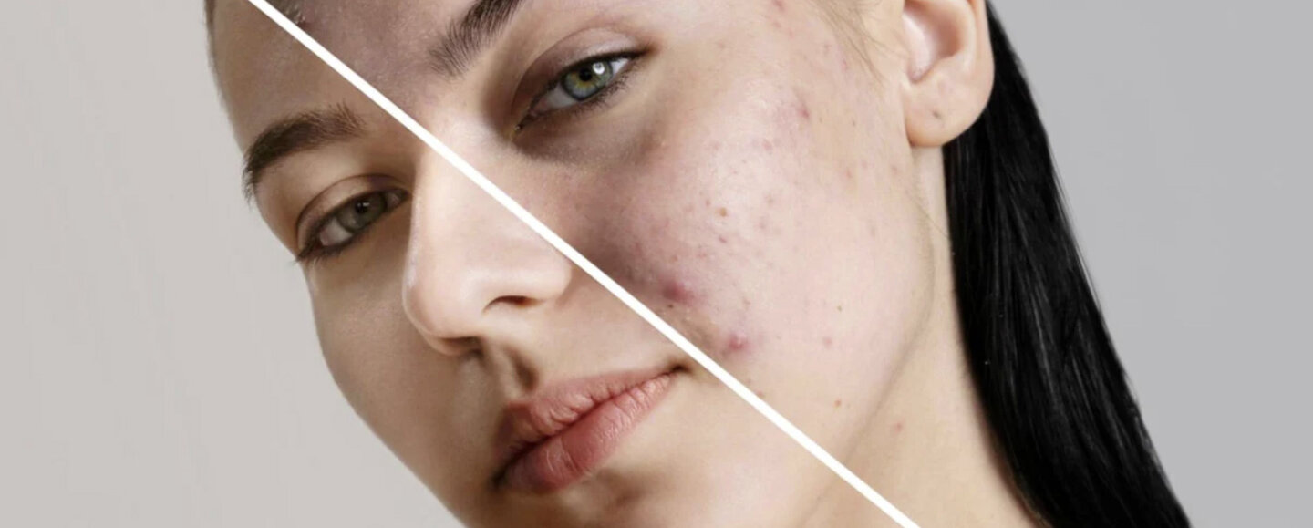
Half of earths population has sensitive skin, however significant variations among countries exists (4). Stinging, itching, burning, tingling, and prickling are sensations familiar to people with sensitive skin. It is defined as a syndrome of appearance of unpleasant sensations in response to chemical or physical stimuli that do not provoke similar sensations in normal skin. These sensations cannot be explained solely by skin disease, irritation, allergy or disruption of barrier function. Often unpleasant sensations are provoked by external (cosmetics, water, household products, pollution, sun exposure, temperature, wind) or internal (emotions, stress, hormones) factors. Interestingly, sensitivity to one trigger cannot predict sensitivity to others.
Sensitive skin can appear normal, without any visible clinical manifestations. It can be accompanied by erythema or not, it can be dry, oily, mixed or normal (7). It affects all body locations, especially face. The face usually remains uncovered, and exposed to environmental stresses, is highly innervated and regularly treated with cosmetics.
This skin type overreacts to stimuli that does not provoke any reaction in normal skin. We should therefore clearly understand the differences between irritated, sensitised and sensitive skin. Skin is sensitised when an allergy is developed. Inflammatory eczematous reactions are typical for sensitised skin, and only allergic individuals are affected. Skin is irritated when we expose it to for example, a detergent. Unpleasant sensations and irritation (erythema) will appear in 100% of cases. Therefore, all individuals are affected.
The actual physiopathology of sensitive skin syndrome is today, more clearly understood. Paradoxically, sensitive skin contains less nerve fibers (6, 8), especially C-fibers, which are responsible for pain, itch and warmth (9). These stimuli activate sensory neuroreceptors (transient receptor potential (TRP) ion channels) in the nerve fibers. Activation of receptors provokes a release of neuropeptides, such as substance P and CGRP (calcitonin-gene related peptide), and thus leads to neurogenic inflammation. Starting from this point, a vicious cycle easily ensues – inflammation activates keratinocytes that secrete more substance P and CGRP amplifying neural sensitisation. Mast cell degranulation as well as deficiency in adiponectin may also play role (10, 11). Sensitive skin is also a neuronal syndrome. The enormous variety of cosmetic products claiming “for sensitive skin” aims to repair a disrupted skin barrier and/or reduce erythema. However, meticulous analysis of published data does not confirm any correlation between sensitive skin syndrome and reduced barrier integrity; the thickness of the epidermis or stratumcorneum; sebum production; pH abnormalities; and skin hydration levels. Furthermore, skin redness is not even necessarily observed. Indeed, some studies show vasoconstriction, while other studies discover vasodilation in sensitive skin (5). Consequently, the mixing neuronal and immunological mechanisms makes it extremely difficult to develop efficient so-called soothing products for sensitive skin. This then results in brands hitting the wrong targets; creating non-efficient cosmetics; misleading their customers; and importantly compromising the industry and ruining the trust of consumers generally.
“
“A study in healthy women providing probiotic yogurt for four weeks showed an improvement in emotional responses as measured by brain scans”
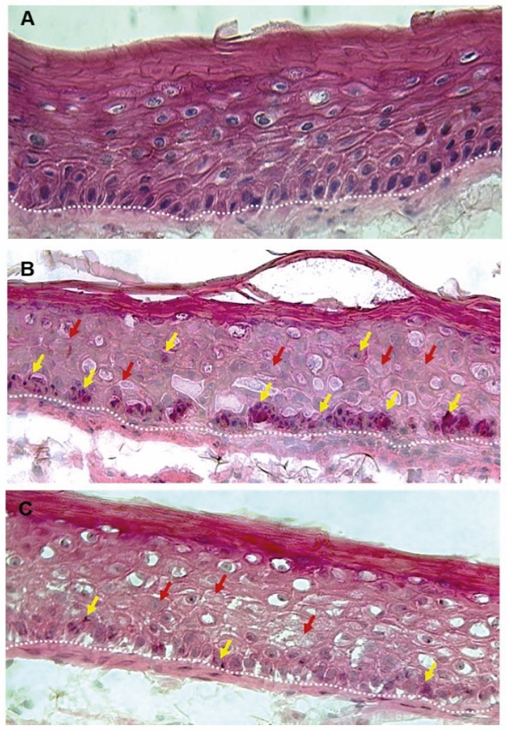
Figure 1. Skin Section with Microbiome. Most microorganisms live in the superficial layers of the stratum corneum and in the upper parts of the hair follicles. Some reside in the deeper areas of the hair follicles and are beyond the reach of ordinary disinfection procedures. There bacteria are a reservoir for recolonization after the surface bacteria are removed.
Materials and methods
Studies of major depressive disorder have been correlated with reduced Lactobacillus and Bifidobacteria and symptom severity has been correlated to changes in Firmicutes, Actinobacteria, and Bacteriodes. Gut microbiota that contain more butyrate producers have been correlated with improved quality of life (1).
A study in healthy women providing probiotic yogurt for four weeks showed an improvement in emotional responses as measured by brain scans (2). A subsequent study by Mohammadi et al. (3) investigated the impacts of probiotic yogurt and probiotic capsules over 6 weeks and found a significant improvement in depression-anxiety-stress scores in subjects taking the specific strains of probiotics contained in the yogurt or capsules. Other studies with probiotics have indicated improvements in depression scores, anxiety, postpartum depression and mood rating in an elderly population (4-7).
Other studies have indicated a benefit of probiotic supplementation in alleviating symptoms of stress. In particular, researchers have looked at stress in students as they prepared for exams, while also evaluating other health indicators such as flu and cold symptoms (1). In healthy people, there is an indication that probiotic supplementation may help to maintain memory function under conditions of acute stress.
Available Tests and Models
There are three models available for tests of soothing cosmetic products, and the choice depends on the stage of the product development process. For pre-clinical tests the researcher may use an in vitro cell culture of human sensory neurons, or a co-culture of human primary keratinocytes and neurons. Soothing activity of a product can be evaluated by biochemical quantification of neuropeptides (substance P) or immunostaining of receptors (TRPV1). This model is poorly adapted for final formulations as the test samples must be soluble in culture medium. A very important note, is that the neuronal monolayer is far from the tridimentional architecture of real human skin with its great diversity of different cellular types.
Overcoming these limits, the researcher may choose a more comprehensive model, comprising re-innervated living human skin explants - ex vivo. The first attempts to develop such model were made several years ago, when rat neurons were co-cultured with human skin explants (12). From scientific point of view this model had some success – animal neurons had successfully created functional nerve endings within the human epidermis. However, this model obviously could not be used for real cosmetic tests due to regulatory constraints. Fortunately, and more recently, an entirely human re-innervated skin, responsive to stimuli, has been developed (13). Currently, pre-clinical tests for soothing products on an ex vivo model mimicking human sensitive skin is available on the market.
Technically, preparation of a re-innervated ex vivo skin model is challenging. At the very first step induced pluripotent stem cells must be isolated from excised human skin biopsies. Then, stem cells are required to be differentiated into functional human sensory neurons, using specific culture medium. Then the co-culture of human neurons, in vitro, with living human skin explants ex vivo, comprises the third step. Furthermore, topical application of a triggering molecule, such as lactic acid or capsaicin on re-innervated explants provokes reactions, similar to those of sensitive skin – secretion of neuropeptides (substance P, CGRP), activation of sensory receptors (TRPV1, TRPA1) and markers of neuroinflammation (TSLP). An efficient soothing product would able to revert these symptoms – reduce secretion of the first, and then decrease activation of the second.
A more detailed publication of the described re-innervated human skin model of sensitive skin, is in progress and will be shortly available in the PubMed database.
The third available model is currently a ‘gold standard’ in efficacy testing for the cosmetic industry. Clinical studies on subjects having sensitive skin can be easily conducted by almost any contract research organization (CRO). The easiest way is to recruit individuals, self-declaring sensitive skin. The stinging test is the most common proposed by CROs in order to evaluate efficacy of a soothing cosmetic product. 10% aqueous solution of lactic acid versus physiological saline is applied topically onto nasolabial folds which provokes unpleasant sensations. Volunteers are required to give a score of the sensation intensity. When the test is repeated after 4 weeks of treatment with a soothing product, this score should (hopefully) be lower. Complementary specific questionnaires may also be used in the clinical study, to collect information regarding product efficacy and/or consumer perceptions about its effects. Regardless, such feedback always provides valuable data. It is important to remember however, that individuals may either be “stingers” or “non-stingers”, and that skin reactive to dry air or sun exposure, may not be sensitive to lactic acid or capsaicin.
The main limit of the stinging test is its lack of objectivity since the given score by the volunteer(s) is absolutely subjective. Objective instrumental measurement is possible when the technique of skin electrodermal response (14) is employed. The principle of the method is based on the well-known polygraph. Detectors to sweat are fixed on fingers, while a thermal electrode is in contact with cheek. Thermal shock stimulates a micro-sweat reaction, which is more important for subjects with sensitive skin. When the test is repeated after 28 days of regular application of a soothing product, the value of the electrodermal response is lower (14).
Balancing
the skin mocrobiome
Studies of major depressive disorder have been correlated with reduced Lactobacillus and Bifidobacteria and symptom severity has been correlated to changes in Firmicutes, Actinobacteria, and Bacteriodes. Gut microbiota that contain more butyrate producers have been correlated with improved quality of life (1).
A study in healthy women providing probiotic yogurt for four weeks showed an improvement in emotional responses as measured by brain scans (2). A subsequent study by Mohammadi et al. (3) investigated the impacts of probiotic yogurt and probiotic capsules over 6 weeks and found a significant improvement in depression-anxiety-stress scores in subjects taking the specific strains of probiotics contained in the yogurt or capsules. Other studies with probiotics have indicated improvements in depression scores, anxiety, postpartum depression and mood rating in an elderly population (4-7).
Other studies have indicated a benefit of probiotic supplementation in alleviating symptoms of stress. In particular, researchers have looked at stress in students as they prepared for exams, while also evaluating other health indicators such as flu and cold symptoms (1). In healthy people, there is an indication that probiotic supplementation may help to maintain memory function under conditions of acute stress.
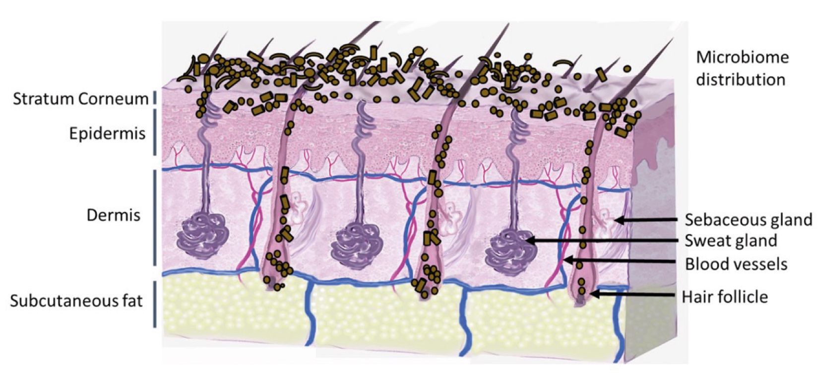
Figure 2. Skin Section with Microbiome. Most microorganisms live in the superficial layers of the stratum corneum and in the upper parts of the hair follicles. Some reside in the deeper areas of the hair follicles and are beyond the reach of ordinary disinfection procedures. There bacteria are a reservoir for recolonization after the surface bacteria are removed.
Conclusion
Sensitive skin is a major concern for more than 60% of women, and nearly 50% of men. Cosmetic and household products are among the main triggers for unpleasant sensations when skin is reactive to them. Sensitive skin is characterized by a decrease in nerve fibers density, hyper-activation of sensory neuroreceptors, and an excessive secretion of neuropeptides. The sequence of these events causes neurogenic inflammation. We should clearly understand not to mistake sensitive with irritated skin, mainly because erythema is not a constant phenomenon of sensitive skin. Understanding the exact mechanisms underlying the hypersensitivity of human skin is crucial for the development of efficient and non-misleading soothing products.
About the Author
Nikita Radionov
Nikita Radionov. Biologist with 8 years of research career. Having PhD in plant physiology, my research experience is based on two post-doctoral positions with particular interest in family of insulin receptors and renal mechanisms of hypertension. Currently I am working as responsible of commercial service at Eurofins BIO-EC.
Nikita Radionov
Head of Sales, PhD. Eurofins BIO-EC, France

References and notes
*This work was presented in part at Cosmetics 360 2022 and InCosmetics Global 2023
- Bernstein ET. Cleansing of sensitive skin; with determination of the pH of the skin following use of soap and a soap substitute. J Invest Dermatol 1947; 9:5—9.
- Misery L, Ständer S, Szepietowski JC, Reich A, Wallengren J, Evers AW, et al. Definition of sensitive skin: an expert position paper from the special interest group on sensitive skin of the international forum for the study of Itch. Acta Derm Venereol. (2017) 97:4–6. doi: 10.2340/00015555-2397
- Misery L, Bataille A, Talagas M, Le Gall-Ianotto C, Fouchard M, Huet F, Ficheux A-S, Roudot A-C, Fluhr JW and Brenaut E (2022) Sensitive Skin Syndrome: A Low-Noise Small-Fiber Neuropathy Related to Environmental Factors? Front. Pain Res. 3:853491. doi: 10.3389/fpain.2022.853491
- Talagas M and Misery L (2019) Role of Keratinocytes in Sensitive Skin. Front. Med. 6:108. doi: 10.3389/fmed.2019.00108
- Richters R, Falcone D, Uzunbajakava N, Verkruysse W, van Erp P, Van de Kerkhof PC. What is sensitive skin? A systematic litera- ture review of objective measurements. Skin Pharmacol Physiol 2015;28:75—83.
- Buhé, V., Vié, K., Guéré, C., Natalizio, A., Lhéritier, C., Le Gall-Ianotto, C., Huet, F., Talagas, M., Lebonvallet, N., Marcorelles, P., Carré, J.-L., & Misery, L. (2015). Pathophysiological Study of Sensitive Skin. Acta Dermato-Venereologica, 96(3), 314–318.
- Misery L, Boussetta S, Nocera T, Perez-Cullell N, Taieb C. Sensitive skin in Europe. J Eur Acad Dermatol Venereol 2009;23:376—81.
- Misery L, Weisshaar E, Brenaut E, Evers AWM, Huet F, Ständer S, et al. Pathophysiology and management of sensitive skin: position paper from the Special interest Group on sensitive skin of the International Forum for the Study of Itch (IFSI). J Eur Acad Dermatol Venereol. (2020) 34:222–9. doi: 10.1111/jdv.16000
- Misery L. Neuropsychiatric factors in sensitive skin. Clin Dermatol. (2017) 35:281–4. doi: 10.1016/j.clindermatol.2017.01.011
- Quatresooz P, Pierard-Franchimont C, Pierard GE. Vulnerability of reactive skin to electric current perception–a pilot study implicating mast cells and the lymphatic microvasculature. J Cosmet Dermatol. (2009) 8:186–9. doi: 10.1111/j.1473-2165.2009. 00445.x
- Kim EJ, Lee DH, Kim YK, Eun HC, Chung JH. Adiponectin deficiency contributes to sensitivity in human skin. J Invest Dermatol. (2015) 135:2331– 234. doi: 10.1038/jid.2015.150
- Lebonvallet N, Boulais N, Le Gall C, Pereira U, Gauché D, Gobin E, et al. Effects of the reinnervation of organotypic skin explants on the epidermis. Exp Dermatol. 2012; 21(2):156-8.
- Lebonvallet N. Re‐innervated skin models of non‐histaminergic itch and skin neurogenic inflammation. Oral presentation at the International symposium by SFC, 6/7 december 2022
- Villaret A, Lestienne F, Vial F, Aubert A, Maret A, El Marbouh L, Bessou-Touya S, Castex-Rizzi N, Duplan H, Coubetergues H. Clinical evaluation of anaesthetic-like effect of two dermocosmetic formulations containing Aquaphilus dolomiae extract-G3 in subjects with sensitive facial skin. J Eur Acad Dermatol Venereol. (2022) Apr;36