Peptides
Skin care
KEYWORDS
PEPTIDES;
SKIN AGING;
NATURAL ACTIVE INGREDIENTS;
Sensor technology;
Detergents;
Sustainability.
peer-reviewed
In vitro investigations of anti-aging benefits provided by natural peptides
Mélanie Coirier, Sylvie Bordes, Elodie Aymard, Hélène Muchico*, Brigitte Closs
*Corresponding author
SILAB, Brive, France
ABSTRACT: Peptides are attracting growing interest in cosmetics for their ability to specifically target cellular mechanisms involved in skin aging, and are especially valued for their capacity to correct its earliest signs. Anti-wrinkles, antioxidant, anti-inflammatory or healing, their mechanisms of action in many physiological processes make peptides attractive for many skin-related indications.
This publication aims to highlight the various anti-aging benefits of natural peptides by exploring the in vitro activity on reconstructed skin models of two specific active ingredients, obtained through eco-extraction processes specific to each raw material.
Introduction
Peptides are small chains of 2 to 50 amino acids linked by peptide bonds (1). As peptide chains form between joining of the primary structure of amino acids, they may enlarge to become oligopeptides, which gather between 10 to 20 amino acids in the chain. Beyond 20 amino acids, the peptide is an unbranched chain named a polypeptide (Figure 1).
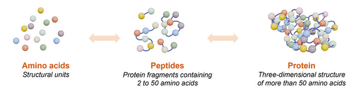
Figure 1. Schematic representation of amino acids, peptides and protein organization.
They have been divided into four main groups, corresponding to their mechanism of action: i) signal peptides, that stimulate matrix protein production, such as collagen or elastin, and cell metabolic functions like cell growth, ii) carrier peptides, iii) neurotransmitter-inhibitor peptides, and iv) enzyme-inhibiting peptides.
Peptides are naturally occurring biological molecules that can be synthesized by all the cells of the organisms. They ensure varied activities required for the correct functioning of the organism, depending on the nature and sequence of their component amino acids (2).
Most peptides on the cosmetics market are synthetics (3). Indeed, peptide synthesis is a technology commonly used to produce peptides of interest with a defined sequence. Peptides are chemically synthesized by the condensation reaction of the carboxyl group of one amino acid to the amino group of another (Figure 2A). While they stand out by their elevated biological specificity, these peptides obtained by chemical processes have limited activity towards a unique biological target. Moreover, if chemical synthesis displays precision tailor made synthesis, it can also impact the environment. At the present time, this lack of transversality and of naturality is no longer consistent with the needs and expectations of consumers who are demanding natural, multifunctional, and effective products (4).
Natural peptides are obtained by the hydrolysis of proteins in small fragments, allowing to expose peptides previously protected inside three-dimensional structures (Figure 2B). There are numerous sources to obtain natural peptides, from plants to microorganisms. Indeed, roots, stem, leaf, flowers, microalgae, yeast, and bacteria all contain proteins. By coupling this diversity of raw materials to enzymes able to hydrolyze proteins, the combination of resulting natural peptides is endless. The choice of the enzyme is based on the nature of the raw material to hydrolyze and the properties of the enzyme. It is also possible to use combinations of enzymes to refine the selection of resulting peptides. The reaction must be stopped at the right time in order to avoid excessive cascade reactions that denature the desired product. This is done by an irreversible thermal inactivation of the enzyme that modifies its active site. The inactivated enzyme is now simply an inert reaction compound that no longer influences the final product. According to standard ISO 16128 concerning cosmetic products, the enzymatic hydrolysis reaction allows to obtain ingredients of natural origin. The resulting diversity of peptides derived from enzymatic hydrolysis enables to achieve specific bio-activity thanks to the unicity of the process.
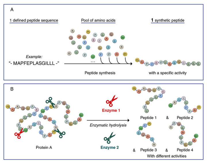
Figure 2. Schematic representation of the obtention of synthetic peptide (A.) and natural peptides (B.). This schematic representation is for illustrative purposes only and does not represent quantitative data.
Cranberry-derived biopeptides for a transversal anti-aging efficacy
Context
The plant kingdom contains an extremely broad diversity of molecules, among which natural peptides, that stand out as very promising active compounds. Although some peptides exist in their free form, most are derived from native proteins from which they are released by hydrolysis. Certain plants are natural protein sources, just as plant co-products from the food processing industry such as press cakes (5). The beneficial use of these co-products also responds to the growing desire of consumers who prioritize cosmetic care products obtained from the botanical world (6).
In this context, SILAB has decided to explore the molecular richness of cranberry fruit press cakes. This superfruit is known for its nutritional value and benefits for health. Enzyme engineering and purification expertise have been beneficially used to offer a peptide concentrate combining richness and performance. The sequential action of two proteases specifically selected has enabled the optimized extraction of all the peptides of interest present in this cranberry co-product. The use of molecular sorting combined with purification by ion exchange chromatography on resin enables the active fraction to subsequently be concentrated in natural peptides particularly rich in arginine (Figure 3). This unique patented process led to the development of the Vaccinium Macrocarpon Fruit Extract.
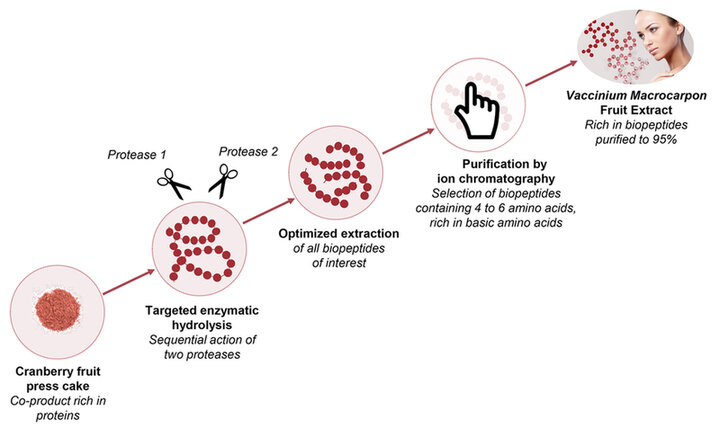
Figure 3. Process adapted to the extraction of natural peptides from cranberry fruit press cakes.
The molecular diversity of the Vaccinium Macrocarpon Fruit Extract favors a transversal biological activity of the active ingredient, as shown by a study of its potential for activity on the major genes involved in cutaneous homeostasis.
Experimental procedure
This study was conducted on 34 genes involved in the different processes related to establishing:
- the physical or chemical epidermal barrier function: proliferation, hydration, cohesion, differentiation, inflammation and antioxidant system;
- the dermal-epidermal junction (DEJ);
- matrix dynamics: matrix organization and degradation, hydration and inflammation.
The expression of these genes was determined by quantitative PCR on human keratinocytes from young (≤ 30 years old) and old donors (≥ 60 years old), and on young human fibroblasts (< P5) and aged fibroblasts obtained by successive passages (> P20). The studies were conducted on four different donors, and each donor was tested twice, which corresponds eight biological replicates. The results correspond to the average value of the -dcT obtained for the eight samples.
A cytotoxicity test using the MTT method was performed to evaluate the effect of the product on cell viability. The cells (keratinocytes and fibroblasts) were exposed to different concentrations of the product, up to 5%. The results show that the product does not induce cytotoxicity, with cell viability remaining above the 90% threshold for all concentrations tested.
Young and old human cells were seeded and incubated at 37°C in an atmosphere containing 5% CO2. After 4 days, the cells were treated with the Vaccinium Macrocarpon Fruit Extract at 0.5% (V/V) or with a 1 µM solution of retinol for 48 hours. After 6 days, cells were recovered and total RNAs were extracted.
RNAs were reverse transcribed, and the complementary DNAs obtained were analyzed with the quantitative PCR technique. The mRNAs of proteins GUSB, RPS18 and GAPDH, internal references standards, were analyzed in parallel to the mRNA of the markers involved in the epidermal barrier function, the DEJ and the dermal matrix.
Fluorescence incorporation (SYBR Green) was quantified continuously with an LC480 LightCycler thermocycler (Roche). Analysis of Ct (relative quantification) was done with LC480 software (Roche).
Statistical analysis was performed with the Student's t test for paired samples (the data follow a normal distribution: Shapiro-Wilk test for normality greater than 5%). In modeling, the results are compared with a two-tailed test and in efficacy with a one-tailed test with, in both cases, a significance level of 5%.
Results
Results demonstrate that among 34 genes evaluated, 76% exhibit an expression that is modified in the course of aging, reflecting a major impact on the barrier function, the DEJ and the dermal matrix.
With the Vaccinium Macrocarpon Fruit Extract Expression the expression of 81% of deregulated cutaneous homeostasis genes is normalized compared to old cells control. Indeed, tested at 0.5% on old keratinocytes, it significantly regulates the expression of genes related to proliferation (Ki-67: +41%), differentiation (loricrin: +75% and filaggrin: +47%), cohesion (claudin-1: +30%), and anti-free radical defense (SOD1: +12%). Tested at 0.5% on old fibroblasts, it also has a significant action on the expression of genes related to matrix organization (collagen I: +24% and elastin: +28%) and its degradation (MMP-1: -37%) (Figure 4).
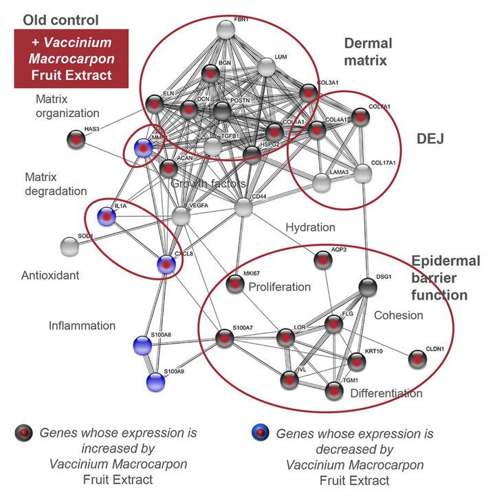
Figure 4. Analysis of the potential of the Vaccinium Macrocarpon Fruit Extract by a targeted transcriptomics analysis.
These results suggest that this anti-aging active ingredient restores homeostasis of mature skin by acting on the dermis, the dermal-epidermal junction and the epidermis.
Rice-derived di- and tripeptides to protect the skin against nutritional deficiencies
Context
Aging involves two biological and clinical distinct processes. Intrinsic aging, in other words biological aging, affects the skin and all organs. Extrinsic aging results from environmental factors, including ultraviolet radiation (UV), smoking, pollution, lack of sleep, but also an unbalanced diet (7). The appearance of the skin is in fact the faithful reflection of health and wellbeing, but also that of deficiencies and poor eating habits. The development of nutraceutics and cosmetofoods has sensitized the cosmetics market to issues of skin nutrition and dietetics. Deficiencies in certain nutrients (vitamins, trace elements, amino acids, essential fatty acids, proteins) reduce the metabolic capacities of skin cells and is the starting point for premature aging of the skin.
The nutrition issue involves not so much the supply of nutrients but rather their bioavailability. In the areas of athletic and infantile dietetics, it is well known that the supply of nutrients that can be rapidly assimilated facilitates metabolic functions, in particular the body's recovery capacities after an effort or a stress. It is known that the size of peptides determines their assimilation by cells and the resulting activity. Considerable work has been done in the areas of artificial nutrition and dietetics of patients incapable of normal ingestion resulting from physical damage or incapacity (accident, surgery, coma, burn victims, etc.) where nutrients, in particular nitrogen as proteins, are directly introduced into the bloodstream. In conditions where the digestion of proteins is not possible, work has shown that proteins in the form of small molecules (primarily di- and tripeptides) are more rapidly absorbed and assimilated than larger peptides or amino acids (8).
Rich in rice di- and tripeptides, the Hydrolyzed Rice Protein Extract was developed to compensate for nutritional imbalances of the skin.
Experimental procedure
In order to assess the capacity of the Hydrolyzed Rice Protein Extract to compensate the deficiency of nutrients (growth factors) required to construct skin, two studies were conducted: one on the formation of an epidermis in a 3D model of nutritional deficiency and one on key proteins of the extracellular matrix and dermal-epidermal junction, also in a 3D model of nutritional deficiency. The studies were conducted on two different donors, and each donor was tested twice. There are therefore four biological replicates.
Study of the formation of an epidermis in a 3D model of nutritional deficiency
Reconstructed skin (RS) was grown in the presence of the Hydrolyzed Rice Protein Extract in complete or deficient culture medium. The efficacy was assessed by measuring morphological modifications of reconstructed skin by hematoxylin-eosin-saffron (HES) staining and by highlighting modifications of the epidermal compartment by immunohistological labeling of Ki-67, integrin α2β1, filaggrin and loricrin.
To this end, human keratinocytes were seeded on lattices containing human fibroblasts and incubated at 37°C in an atmosphere containing 5% CO2. On days 2, 4, 7 and 9, the culture medium was discarded and replaced with complete medium or medium depleted in growth factors and containing the Hydrolyzed Rice Protein Extract at 0.10% or 0.25%. Reconstructed skin samples were then incubated at 37°C in an atmosphere containing 5% CO2. After 11 days, reconstructed skin was fixed, dehydrated and embedded in paraffin for histology studies. A Leica RM2125RT microtome was used to prepare 4 µm sections.
The morphological analysis of reconstructed skin by HES staining and the immunohistofluorescence analysis were both done with an IX 70 microscope (Olympus) coupled to an image analysis system (NIS-Elements AR, Nikon). Epidermal thickness (layers of living cells) was measured on the histology sections after HES staining. As for the synthesis of different markers examined, it is proportional to the intensity of green fluorescence in reconstructed skin samples. Quantitative image analysis was conducted with Matlab® software, version R2012b (MathWorks).
Study of key proteins of the extracellular matrix and dermal-epidermal junction in a 3D model of nutritional deficiency
Reconstructed skin was grown in the presence of the Hydrolyzed Rice Protein Extract in culture medium depleted to varying extents. The efficacy of the product was examined by analyzing the synthesis of procollagen I by ELISA assay, of fibrillin-1 (a marker of the dermal matrix) and of collagen VII (a marker of the dermal-epidermal junction) by immunohistological labeling.
The same previous-mentioned protocol was applied, except on day 11, when culture media were recovered and frozen at -80°C before assay. For histology examination, reconstructed skin samples were embedded in Tissue-Tek® and frozen. 4 µm sections were then prepared with a Leica CM1850 cryostat.
Procollagen I was assayed using an ELISA kit (procollagen type I, Takara). The immunohistofluorescence analysis of fibrillin-1 and collagen VII was done with an IX 70 microscope (Olympus) coupled to an image analysis system (NIS-Elements AR, Nikon). The synthesis of different markers examined is proportional to the intensity of green fluorescence in reconstructed skin samples. Quantitative image analysis was conducted with Matlab® software, version R2012b (MathWorks).
Results
Study of the formation of an epidermis in a 3D model of nutritional deficiency
Results show that, tested at 0.25%, the Hydrolyzed Rice Protein Extract significantly increases thickness of the epidermis by 19% in normal reconstructed skin and significantly returns thickness of the epidermis to normal in deficient reconstructed skin (+91%).
In addition, the immunohistofluorescence analysis shows that the Hydrolyzed Rice Protein Extract significantly increases epidermal markers in normal reconstructed skin (Ki-67: +34%, Integrin α2β1: +34%, loricrin: +29%, filaggrin: +32%) (Figure 5).
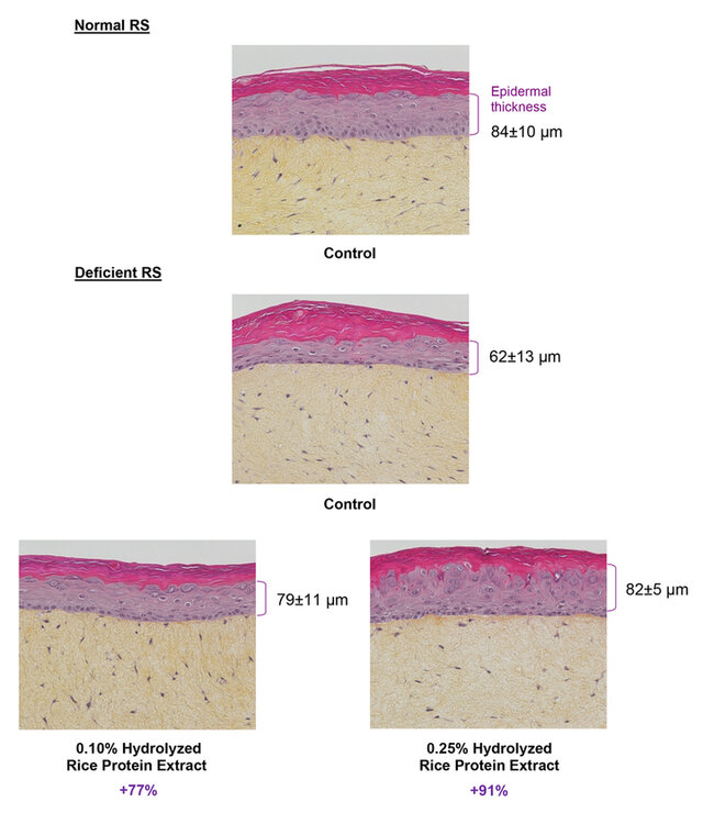
Figure 5. Effect of the Hydrolyzed Rice Protein Extract on the thickness of the epidermal compartment of deficient reconstructed skin.
Study of key proteins of the extracellular matrix and dermal-epidermal junction in a 3D model of nutritional deficiency
Tested at 0.25%, the Hydrolyzed Rice Protein Extract significantly increases the synthesis of procollagen I by 17% in normal reconstructed skin as well as the synthesis of procollagen I by 37% in deficient reconstructed skin.
The active ingredient also significantly increases the synthesis of fibrillin-1 by 28% in normal reconstructed skin and returns it to normal (+150%) in deficient reconstructed skin.
Last, it significantly boosts the synthesis of collagen VII by 14% in normal reconstructed skin and maintains the synthesis of collagen VII at 80% of normal in deficient reconstructed skin (Figure 6).
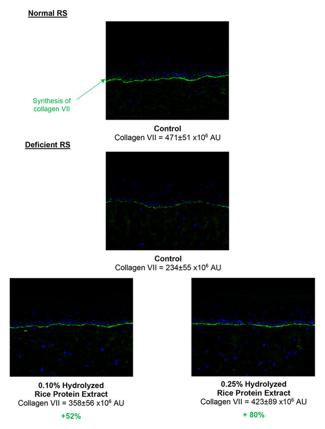
Figure 6. Effect of the Hydrolyzed Rice Protein Extract on the synthesis of collagen VII by deficient reconstructed skin.
All these results show that the Hydrolyzed Rice Protein Extract re-establishes the expression of markers of the extracellular matrix and the dermal-epidermal junction and also enables the reconstruction of mature epidermis.
Conclusion
For its Vaccinium Macrocarpon Fruit Extract, the company has identified an overall anti-aging potential in the cranberry co-product resulting from its high protein content. A technological process was then developed to extract and purify these biopeptides. The powerful anti-aging efficacy of this active ingredient results from the molecular diversity of cranberry biopeptides. It favors the proliferation of keratinocytes and their differentiation, thereby strengthening the barrier function and giving a healthy-glow effect. In addition, the active ingredient restores the undulated structure of the dermal-epidermal junction and boosts the synthesis of matrix components, thereby providing an anti-wrinkle action.
The Hydrolyzed Rice Protein Extract is rich in rice di- and tripeptides. It revitalizes metabolic functions and improves recovery capacities after stress by supplying the skin with nutrients required for its correct operation and functioning (data not shown in this publication). It compensates for nutritional imbalances by maintaining the synthesis of dermal-epidermal junction proteins and extracellular matrix proteins, and also by enabling the formation of a mature epidermis. With this ingredient, the skin is better nourished and revitalized, and once again has its youthful appearance.
About the Author
Hélène MUCHICO is Marketing and Communication manager of SILAB, a French-based company engineering natural active ingredients for skin and hair care applications. Holder of both an engineer degree in agronomy and a marketing executive certification from HEC Paris, she has been involved in the cosmetic industry for the last 20 years willing to emphasize the power and relevance of natural active ingredients.
Hélène Muchico
SILAB, Brive, France

References and notes
- Forbes J, Krishnamurthy K. Biochemistry, Peptide. 2023 Aug 28. In: StatPearls [Internet]. Treasure Island (FL): StatPearls Publishing; 2025 Jan–. PMID: 32965931. https://pubmed.ncbi.nlm.nih.gov/32965931/
- Zhang L, Falla TJ. Cosmeceuticals and peptides. Clin Dermatol. 2009 Sep-Oct;27(5):485-94. doi: 10.1016/j.clindermatol.2009.05.013. PMID: 19695481. https://linkinghub.elsevier.com/retrieve/pii/S0738081X0900128X
- Ferreira MS, Magalhães MC, Sousa-Lobo JM, Almeida IF. Trending Anti-Aging Peptides. Cosmetics. 2020; 7(4):91. https://doi.org/10.3390/cosmetics7040091. https://www.mdpi.com/2079-9284/7/4/91
- Mintel
- Montesano, D.; Gallo, M.; Blasi, F.; Cossignani, L. (2020) Biopeptides from vegetable proteins: new scientific evidences. In : Current Opinion in Food Science, vol. 31, p. 31–37. https://www.sciencedirect.com/science/article/abs/pii/S2214799319300840
- Real Simple, 13 Top Beauty Trends for 2021. Skin Inc.https://www.skininc.com/business/trends/news/21886647/13-beauty-trends-for-2021
- Schagen SK, Zampeli VA, Makrantonaki E, Zouboulis CC. Discovering the link between nutrition and skin aging. Dermatoendocrinol. 2012 Jul 1;4(3):298-307. doi: 10.4161/derm.22876. PMID: 23467449; PMCID: PMC3583891. https://pmc.ncbi.nlm.nih.gov/articles/PMC3583891/
- Fürst P. The role of amino acids and synthetic dipeptides. Clin Nutr. 2003;22 Suppl 2:S23-8. doi: 10.1016/s0261-5614(03)00147-x. PMID: 14512048. https://pubmed.ncbi.nlm.nih.gov/14512048/