Skin care
Skin care
peer-reviewed
A novel biotechnological active ingredient that enhances electrogenic bacteria to protect skin from oxidative damage and aging
CATRIN YOUSSIF*, CRISTINA BONELL, MAURICIO VALERIO-SANTIAGO, JORDI GÁLVEZ, ALBERT SOLEY,
GEMMA MOLA, RAQUEL DELGADO
*Corresponding author
Lipotec SAU, Gavà (Barcelona), Spain
ABSTRACT:The role of skin microbiota in protecting against aging and photoaging is an emerging concept. Beneficial probiotic bacteria on the skin contribute to mechanisms that help in preventing oxidative stress, which is a key contributor to aging mechanisms. The research explores how the skin’s natural electricity levels produced by bacteria can reduce oxidative damage and cellular senescence, which are linked to collagen degradation and skin aging. We also describe a novel Lactobacillus sp. ferment extract, a biotechnological active ingredient which boosts bioelectricity production by the commensal skin microbiota for rejuvenation purposes. In this sense, boosting the antioxidant mechanisms in Staphylococcus epidermidis(S. epidermidis) is proposed as a novel strategy to improve skin health and mitigate the effects of aging and UV damage. Further clinical studies validated these findings contributing to flawless-looking glass skin with anti-aging skin efficacy.
??????????????????
“
“A study in healthy women providing probiotic yogurt for four weeks showed an improvement in emotional responses as measured by brain scans”
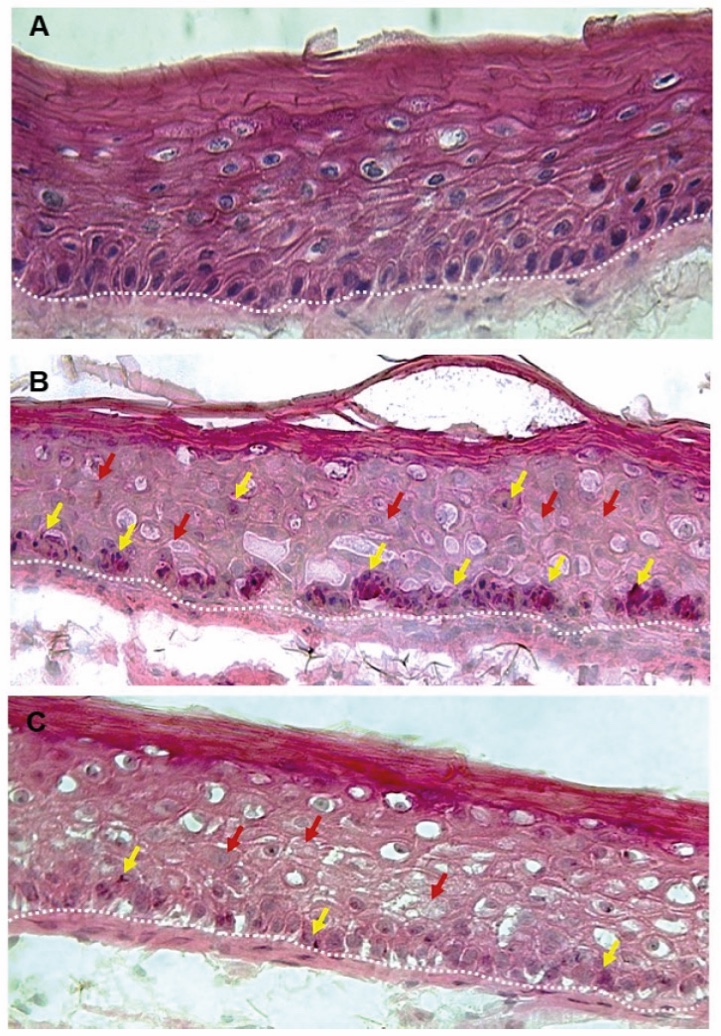
Figure 1. Skin Section with Microbiome. Most microorganisms live in the superficial layers of the stratum corneum and in the upper parts of the hair follicles. Some reside in the deeper areas of the hair follicles and are beyond the reach of ordinary disinfection procedures. There bacteria are a reservoir for recolonization after the surface bacteria are removed.
Materials and methods
Studies of major depressive disorder have been correlated with reduced Lactobacillus and Bifidobacteria and symptom severity has been correlated to changes in Firmicutes, Actinobacteria, and Bacteriodes. Gut microbiota that contain more butyrate producers have been correlated with improved quality of life (1).
A study in healthy women providing probiotic yogurt for four weeks showed an improvement in emotional responses as measured by brain scans (2). A subsequent study by Mohammadi et al. (3) investigated the impacts of probiotic yogurt and probiotic capsules over 6 weeks and found a significant improvement in depression-anxiety-stress scores in subjects taking the specific strains of probiotics contained in the yogurt or capsules. Other studies with probiotics have indicated improvements in depression scores, anxiety, postpartum depression and mood rating in an elderly population (4-7).
Other studies have indicated a benefit of probiotic supplementation in alleviating symptoms of stress. In particular, researchers have looked at stress in students as they prepared for exams, while also evaluating other health indicators such as flu and cold symptoms (1). In healthy people, there is an indication that probiotic supplementation may help to maintain memory function under conditions of acute stress.
Introduction
Recent discoveries have identified bacteria in the gut microbiome that produce electricity using electron transport proteins. These bacteria, known as electrogenic bacteria (1), have also been found on the skin. S. epidermidis is one such bacterium, notable for its importance and prevalence in the human skin microbiome (2). In the skin, S. epidermidis is already known to inhibit the growth of pathogenic bacteria, modulate immune responses, and promote wound healing (3, 4, 5, 6, 7). This newly identified functionality of S. epidermidis leads to a deeper understanding of its role in maintaining skin homeostasis.
Electrogenic bacteria, like S. epidermidis, generate bioelectricity by metabolizing short-chain fatty acids (SCFAs), such as acetic acid, isobutyric acid, isovaleric acid, and methylbutyric acid, that are found on the skin (8). Electrogenic bacteria oxidize the SCFAs and thereby, electrons are generated. In other words, electricity is produced. These electrons are available to neutralize free radicals and mitigate oxidative stress, even when exacerbated by external factors such as UV radiation, minimizing premature aging (9, 10 ,11).
Oxidative stress and cellular senescence are closely linked to aging. Senescent exhibit characteristic changes such as enlarged morphology and increased secretion of pro-inflammatory cytokines and ROS (4, 10, 11, 12, 13). They accumulate with age and contribute to tissue dysfunction and aging. In this sense, ROS-induced and ROS-associated senescence can create a feedback loop, amplifying cellular damage and the senescence phenotype, contributing to the aging process (14, 15). Therefore, combating oxidative stress plays a significant role in skin aging and damage (16, 17, 18, 19, 20, 21, 22). Understanding the interplay between ROS and cellular aging is crucial for developing anti-aging ingredients.
Antioxidants in skincare are widely recognized for their ability to neutralize free radicals and reduce signs of premature aging (23). Moreover, they help reduce collagen degradation induced by ROS (24, 25, 26, 27). Enhancing antioxidant mechanisms in S. epidermidis through the production of bioelectricity may offer new avenues for skin protection, emphasizing the importance of supporting beneficial bacteria to enhance skin resilience, while also providing anti-aging benefits (10, 28, 29, 30, 31).
To achieve this, a novel Lactobacillusextract with a prebiotic effect was produced to enhance the antioxidant mechanism discovered in S. epidermidis. This mechanism involves inducing bioelectricity production in these bacteria (2). This research aims to determine if enhancing this novel mechanism can prevent or reverse damage caused by oxidative stress. Therefore, we first demonstrated the induction of electrogenic, and antioxidant activity induced in S. epidermidis with the active ingredient in vitro. Then, we proved that the application of the biotechnological ferment extract acts as a protective shield against ROS and mitigates signs of aging ex vivo. Finally, further clinical studies have validated the use of this active ingredient in reducing the physical signs of aging and improving overall skin health and appearance.
Materials and methods
Obtention of the bacterial ferment
Lactobacillus sp. ferment (LectroglazeTM biotech ingredient, Lipotec S.A.U., Lubrizol Life Science, Beauty) was developed from a Lactobacillus strain isolated from fermented beets. The biotechnological process to produce the material involved in the fermentation in stirred tank bioreactors, followed by clarification to remove biomass. Ferment broth contains Oligosaccharides (fructo and galactooligosaccharides) (1-3 KDa), lactic acid, and peptides (0.5-1.4 KDa).
Unless otherwise stated, all statistical analysis in vitro were calculated using an unpaired Student’s t-test, and in vivo, a Student’s t-test or Wilcoxon test after checking the normality distributions by Shapiro-Wilk test.
Inducing SCFAs production, electricity and antioxidant capacity of S. epidermidisin vitro.
SCFAs production was measured in aculture containing S. epidermidis treated with Lactobacillus extract for 17h. Acetic acid, isobutyric acid, isovaleric acid and methylbutyric acid were measured by means of a Shimadzu gas-chromatograph equipped with Flame-ionization detector, comparing each analyte to a known control (Scharlab, Sigma-Aldrich) (32,33).
For measuring the potential of theextract to induce electricity generation in S. epidermidis, the bacteria were incubated in the presence/absence of the extract for 24h. Electricity produced by bacteria was detected by using a chamber equipped with a cathode and an anode. The cathode has wrapped up to a Nafion membrane which served as a proton exchange membrane. Voltage changes were recorded by a digital multimeter (Lutron) for 300 seconds. The highest voltage value was used for quantification.
To evaluate the antioxidant activity, S. epidermidis was treated with Lactobacillus extract for 24h. Culture medium alone and with the bacteria were used as basal control. The antioxidant efficacy was evaluated by FRAP (Ferric reducing antioxidant power) assay.
Inhibition of collagen degradation in a skin biopsy model for stress-induced premature senescence (SIPS) in the presence of S. epidermidis.
The rejuvenating effect of Lactobacillus extract was tested ex vivo on human skin explants, in which the SIPS phenotype was induced in the presence of S. epidermidis. Biopsies were treated on the surface with S. epidermidis (4h) and then H2O2 (2mM) was added to the culture medium (2h, systemically). Lactobacillusextract was then applied systemically and topically (24h) daily for 3 days (day 4=collection). Sections were incubated with collagen hybridizing peptide (CHP) Cy3-Conjugate for quantification of denatured collagen by measuring fluorescence intensity.
Clinical evaluation of a cream containing 2% of Lactobacillus extract for the assessment of youthful looking smooth and glowing skin.
A clinical test was carried out on 161 female and male volunteers from Spain and China. The volunteers from Spain were comprised of 95 subjects which were split into a younger group (20-30 y.o.) and an older group (40-60 y.o.). The volunteers from China were comprised of 66 older volunteers only (40-60 y.o). All the volunteers applied either a cream containing 2% of the active ingredient or a placebo cream on the whole face, twice a day for 28 days. After the treatment, pores changes were evaluated.
Specific inclusion criteria:
Age: between 20 and 60 years old (±1 year).
Skin type: Phototype II–III.
Subjects showing pores on cheek area.
Subjects showing light skin tone (Individual Typology Angle > 35).
Evaluation of pores
The number of total pores and the pores index (accounting for pore depth, size and density) were evaluated on the cheek area with Antera 3D® system (Miravex). With age, pores change in shape and become enlarged and elongated, which is mainly caused by the reduced skin collagen and elasticity due to aging (34). The length and area of the age-related pores were analyzed using computer vision and were compared between younger and older groups of volunteers. Briefly, from the images obtained, circular pores were subtracted obtaining an image where only the elongated pores could be analyzed. A second filter for pore size was applied to ensure that only visible age-related pores remained. For each image, the mean length and the percentage of area covered by elongated aged pores were measured.
Another clinical study was conducted with 95 male and female participants from Spain, following the same protocol and assessment criteria as described in the preceding study. After 28 days of treatment, changes in skin translucency, luminosity and hydration were evaluated.
Measurement of skin translucency
Skin translucency was evaluated by measuring the diffuse light scattered within the skin through Translucency Meter TLS (Dia-Stron). A skin translucency 3D map was generated by creating a surface plot with a colormap based on the average measurement values obtained on the cheek from all the volunteers.
Skin luminosity assessment
The luminosity of the skin was evaluated on the cheek through the L* parameter in the cieLab color scale using Antera 3D® system (Miravex).
Measurement of skin hydration
Skin moisturization was assessed using a Corneometer® CM 825 (Courage & Khazaka). A 3D skin hydration map was generated by creating a surface plot with a colormap, based on the average values measured at 5 distinct points on one hemi-face of each volunteer.
Results and discussion
Inducing SCFAs production, electricity and antioxidant capacity of S. epidermidisin vitro.
The capacity of Lactobacillus extract to induce SCFAs in S. epidermidis was demonstrated in vitro, as SCFAs act as electron donors and are thereby able to promote electricity production (5,6,7).
As shown in Figure 1, treatment with Lactobacillus extract induced acetic acid, isobutyric acid, isovaleric acid and methylbutyric acid release in vitro in the presence of S. epidermidis.
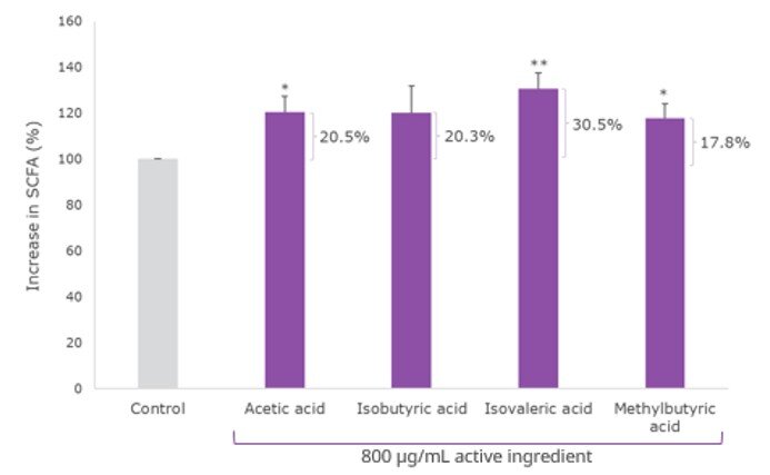
Figure 1. % Induction of SCFAs release measured by HPLC (vs. Control: *p < 0.05; **p < 0.01).
As it is hypothesized that SCFAs production contribute to electricity generation by acting as electron donors (1,2), the capacity of the ingredient to induce electrogenic activity in the bacteria was next evaluated. A significant increase in the levels of electricity production of the S. epidermidis culture in the presence of the Lactobacillus extract was observed compared to the control (Figure 2).
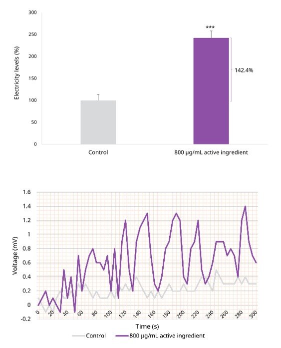
Figure 2. A) % Highest point of electricity in the presence of S. epidermidis (vs Control: *** p < 0.001). B) Representative image showing the changes in electricity along time measured by a digital multimeter.
As electricity may play a key role in neutralizing ROS (7, 9), we evaluated if the Lactobacillus extract enhances bacterial antioxidant activity. As shown in Figure 3, the active ingredient exhibited some antioxidant efficacy on its own. However, when combined with S. epidermidis, the induction of the antioxidant capacity was superior compared to the antioxidant efficacy of either the active ingredient or S. epidermidis alone.
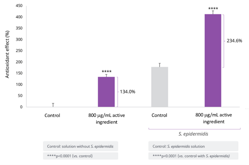
Figure 3. % Antioxidant efficacy measured by FRAP analysis (vs. Control: **** p < 0.0001; vs. Control with S. epidermidis: **** p < 0.0001).
Inhibition of collagen degradation in a skin biopsy model for SIPS in the presence of S. epidermidis.
Lactobacillus extract was able to significantly reduce SIPS induced collagen degradation, minimizing the age-induced collagen disruption for a rejuvenating effect (Figure 4).
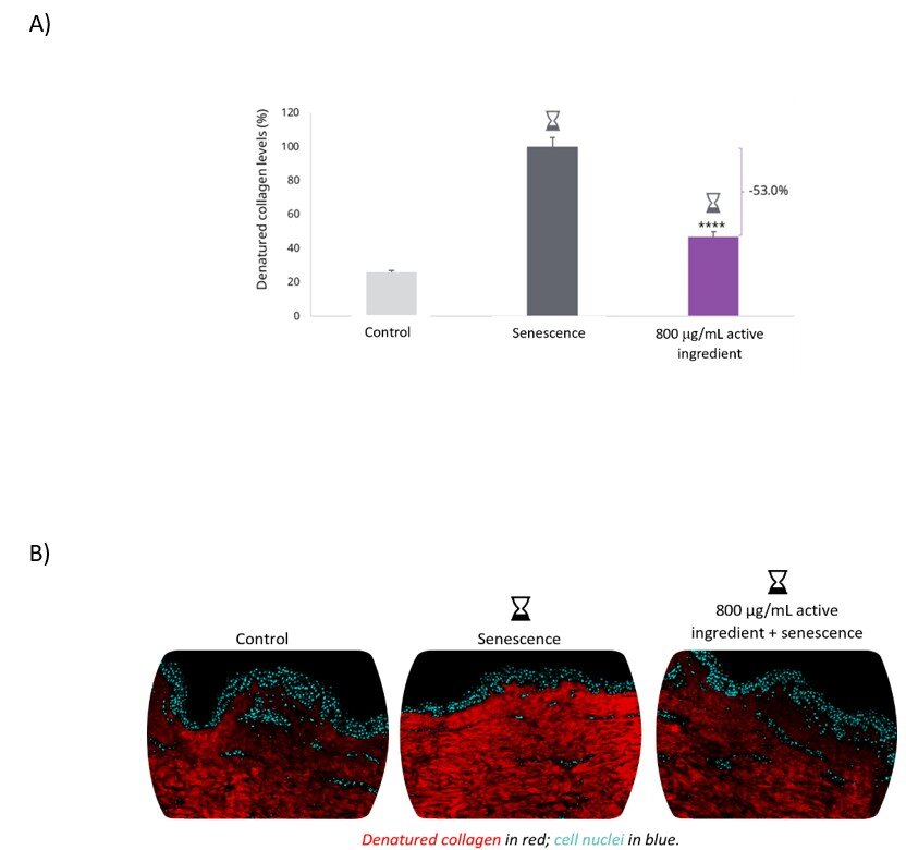
Figure 4. A) % Denatured collagen levels (vs. Senescence: **** p < 0.0001). B) Representative images of collagen degradation in human skin biopsies measured by fluorescence measurement of CHP binding.
Taken together, these results demonstrate that Lactobacillus extract increases SCFAs production in S. epidermidis, thus inducing the generation of bioelectricity and promoting an antioxidant effect to effectively protect the skin from damage caused by oxidative stress.
Clinical efficacy of a 2% Lactobacillus extract cream for the assessment of youthful looking smooth and glowing skin.
Reducing the appearance of pores and age-related pores.
Pore assessment showed a significant decrease in the number of pores and pores index (which accounts for pore depth, size, and density) for smoother skin (Figure 5) after the application of the cream containing 2% of Lactobacillus extract.
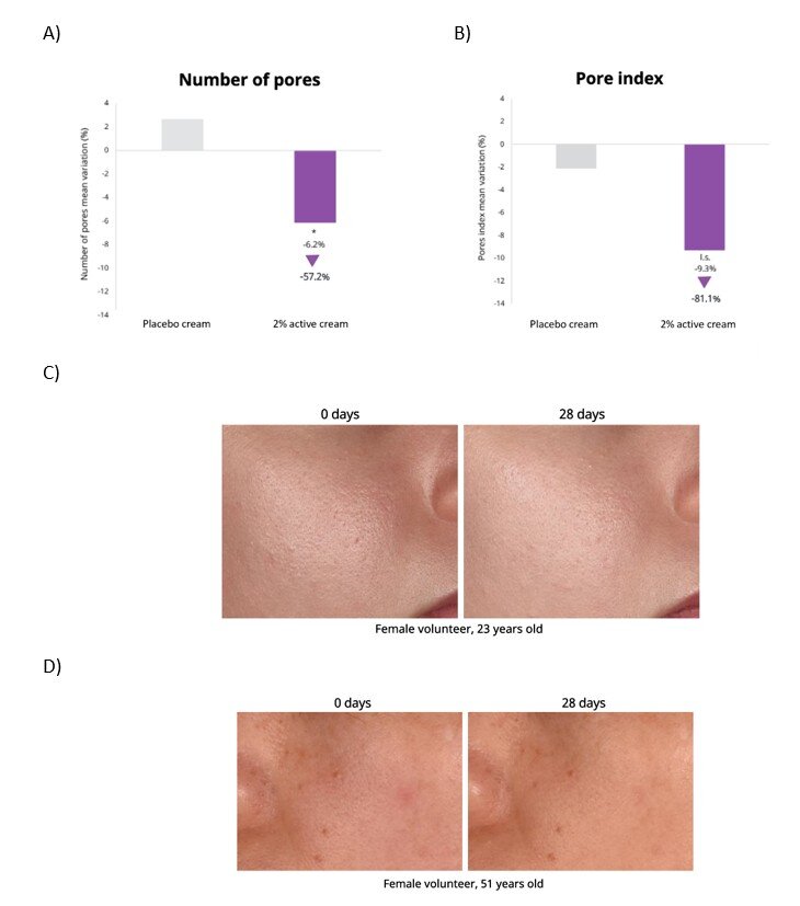
Figure 5. Variation (%) in the: A) Number of pores (*p<0.05 vs. initial time and placebo) B) Pores index (low significance 0.05<p<0.1 vs. initial time on the cheek area using the Antera 3D® system (Miravex). C) Representative images showing minimized appearance of pores in a volunteer from Spainand D) from China.
We then analyzed pore length and area finding an 80% higher prevalence of age-related pores in the older group compared to the younger group (data not shown). Further, the effect of the active cream treatment on the pore’s phenotype was analyzed, showing a significant reduction in elongation, contributing to a younger looking complexion (Figure 6).
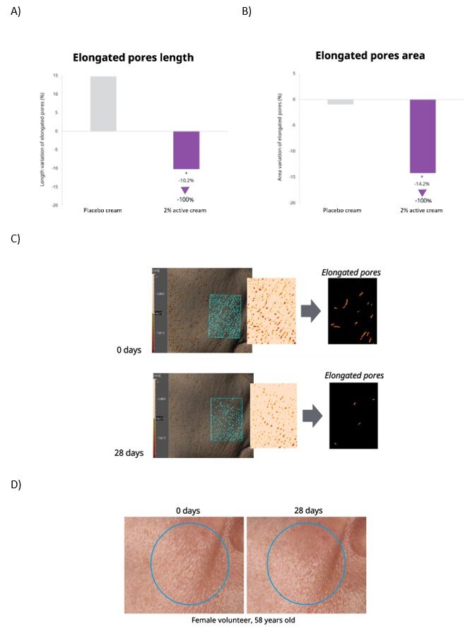
Figure 6. Variation (%) in the A) length of elongated pores (*p<0.05 vs. initial time and placebo), and B) the pore area of elongated pores (*p<0.05 vs. initial time) on the cheek area. C) Representative example of image processing data analysis using Antera 3D® system (Miravex), in which age-related pores were identified by detecting elongated and enlarged pores through computer vision techniques D) Representative images showing the minimized appearance of enlarged pores in a volunteer from Spain.
Increasing skin translucency.
Skin translucency was evaluated, and the results showed a significant increase contributing to a radiant skin complexion and a glow from within effect (Figure 7) after application of the active cream.
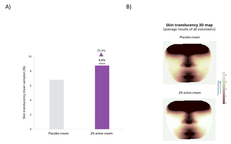
Figure 7. A) Variation (%) in skin translucency (****p<0.0001 vs. initial time) on the cheek area B) Representation of the 3D map of skin translucency generated from the average measurement values of all volunteers using the Translucency Meter TLS (Dia-Stron).
A significant increase in skin luminosity contributing to a glowing skin complexion was observed (Figure 8, 9).
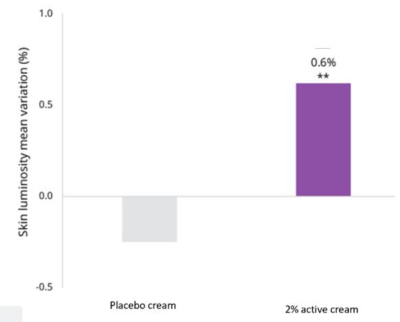
Figure 8. Variation (%) in skin luminosity (**p<0.01 vs. initial time and placebo)evaluated on the cheek area through the L* parameter in the cieLab color scale using Antera 3D® system (Miravex).
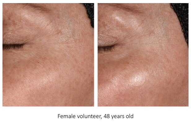
Figure 9. Representative images showing an increase in skin glow in female volunteer from Spain.
Increase in skin hydration
Hydration is also key to achieving natural glow. When skin is dehydrated, it can appear dull and dry,
losing radiance (35). A skin hydration 3D map was generated based on the measurement values obtained at different regions of the face. A significant increase in hydration was observed on the cheek area (Figure 10) after active cream application.
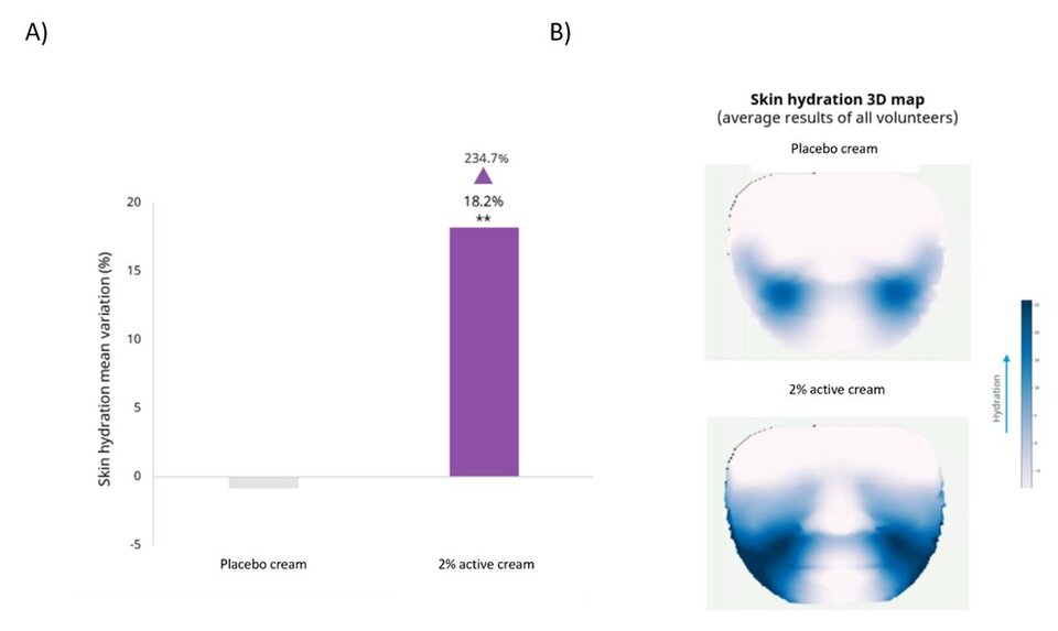
Figure 10. A) Variation (%) in skin hydration (**p<0.01 vs. initial time and *p<0.05 vs. placebo) on the cheek area B) Representation of the 3D map of skin hydration generated from the average measurement values of all volunteers using the Corneometer® CM 825 (Courage&Khazaka).
Conclusions
The novel Lactobacillus extract emerges as an innovative and comprehensive solution to reduce the visibility of age-related pores and improve dull and dry skin. Inspired by the functionality of electrogenic bacteria, a novel active ingredient has been developed to boost the skin’s natural electricity levels, acting as ROS scavengers and thereby offering a significant antioxidant effect. In vitro and ex vivo studies demonstrated the ability of the Lactobacillus extract to protect skin cells from the damaging effects of cellular senescence, helping also to minimize oxidative stress and collagen degradation under these conditions. On volunteers, a cream containing 2% of Lactobacillus extract helped obtain a glowy skin appearance by improving skin translucency, luminosity and hydration, as well as by decreasing the appearance of all pores and of the more specific age‑related pores.
The development of skincare products targeting skin microbiota represents a paradigm shift in the cosmetic field. This approach moves away from conventional methods focused solely on surface‑level concerns to more holistic strategies addressing the underlying causes of skin aging and damage. By leveraging the natural symbiotic relationship between the skin and its resident bacteria, this innovative solution has the potential to revolutionize skincare, promoting healthier, more resilient skin.
Incorporating the Lactobacillus extract into anti-aging skin cosmetic formulations will allow brands and manufacturers to offer consumers a powerful solution that promotes overall skin and microbiome health, combating the aging process by minimizing age-related changes in pore visibility, glow, and hydration.
Conclusion
The future of cosmetics lies in the continued evolution of holistic approaches which represents a transformative shift in the industry, merging scientific advancements, natural ingredients, and wellness principles. By understanding and embracing the interconnectedness of these elements, the cosmetics industry can cultivate products that not only enhance external beauty but also contribute to the overall well-being of individuals and the planet.
The interplay between beauty from within and topical cosmetics is the key for future products. The integration of biotechnology and green chemistry is revolutionizing cosmetic formulations, offering sustainable and biocompatible alternatives.
Developers can implement blockchain to trace the journey of ingredients from source to product. Nevertheless, the efficacy of the natural products should be scientifically proven. Marketers can communicate transparency as a brand value, and parallelly educate consumers by highlighting how specific ingredients contribute to radiant and healthy skin.
By embracing the synergy between these approaches and leveraging scientific advancements, the cosmetics industry can provide consumers with comprehensive beauty solutions that cater to both internal and external dimensions of beauty.
Surfactant Applications
The application area lends itself particularly well to the use of AI. Active today in this area is the US company Potion AI (6). The company provides AI-powered formulation tools for beauty and personal care R&D. Their offerings include Potion GPT, next generation ingredient and formula databases and AI document processing. Potion’s work could have a significant impact on the entire surfactant value chain, from raw material suppliers to end consumers. By using their GPT technology, they can help target work toward novel surfactant molecules that have optimal properties for specific applications. By using their ingredient and formula databases, they can access and analyze a vast amount of data on surfactant performance, safety, and sustainability. By using their AI document processing, they can extract and organize relevant information from patents, scientific papers, and regulatory documents. These capabilities could enable Potion AI's customers to design and optimize surfactant formulations that are more effective, eco-friendly, and cost-efficient. A particularly interesting application for this type of capability is deformulation.
Deformulation is the process of reverse engineering a product's formulation by identifying and quantifying its ingredients. Deformulation can be used for various purposes, such as quality control, competitive analysis, patent infringement, or product improvement. However, deformulation can be challenging, time-consuming, and costly, as it requires sophisticated analytical techniques, expert knowledge, and access to large databases of ingredients and formulas.
AI can potentially enhance and simplify the deformulation process by using data-driven methods to infer the composition and structure of a product from its properties and performance. For example, AI can use machine learning to learn the relationships between ingredients and their effects on the product's characteristics, such as color, texture, fragrance, stability, or efficacy. AI can also use natural language processing to extract and analyze information from various sources, such as labels, patents, literature, or online reviews, to identify the possible ingredients and their concentrations in a product.
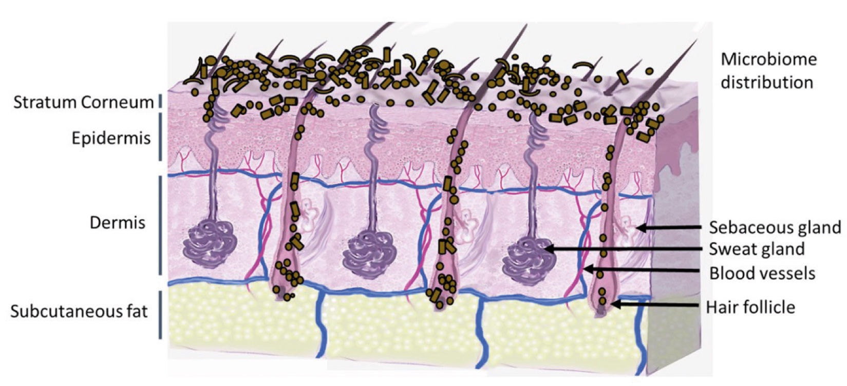
Figure 2. Skin Section with Microbiome. Most microorganisms live in the superficial layers of the stratum corneum and in the upper parts of the hair follicles. Some reside in the deeper areas of the hair follicles and are beyond the reach of ordinary disinfection procedures. There bacteria are a reservoir for recolonization after the surface bacteria are removed.
About the Author
Catrin Youssif
Catrin Youssif holds a master’s degree in Biomedicine and Biotechnology from the University of Cambridge and the University of Veterinary Medicine, Vienna and a PhD degree in Biomedicine from the University of Barcelona performed at the Institute of Biomedical Research Barcelona. She joined Lubrizol in 2018 as an Open Innovation Scientist and, in 2021, advanced to the role of Project Leader in claim substantiation for cosmetic active ingredients.
In her current role, Catrin leads and oversees research activities focused on evaluating the in vitro and in vivo efficacy of new products. Her work involves identifying novel testing methods and molecular pathways, as well as developing innovative solutions that align with evolving market needs. Beyond her scientific responsibilities, she actively contributes to business development initiatives and provides expert scientific support to both commercial and marketing teams.
Catrin Youssif
Lubrizol, Gavà (Barcelona), Spain

References and notes
- Wang, W.; Du, Y.; Yang, S.; Du, X.; Li, M.; Lin, B.; Zhou, J.; Lin, L.; Song, Y.; Li, J. Bacterial Extracellular Electron Transfer Occurs in Mammalian Gut. Anal. Chem. 2019, 91, 12138–12141.https://pubmed.ncbi.nlm.nih.gov/31512863/
 Balasubramaniam A, Adi P, Do Thi TM, Yang JH, Labibah AS, Huang CM. Skin Bacteria Mediate Glycerol Fermentation to Produce Electricity and Resist UV-B. Microorganisms. 2020 Jul 21;8(7):1092.https://pubmed.ncbi.nlm.nih.gov/32708352/
- Byrd AL, Belkaid Y, Segre JA. The human skin microbiome. Nat Rev Microbiol. 2018 Mar;16(3):143-155. https://pubmed.ncbi.nlm.nih.gov/29332945/
- Schommer NN, Gallo RL. Structure and function of the human skin microbiome. Trends Microbiol. 2013 Dec;21(12):660-8. https://pubmed.ncbi.nlm.nih.gov/24238601/
- Manos J. The human microbiome in disease and pathology. APMIS. 2022 Dec;130(12):690-705. https://pubmed.ncbi.nlm.nih.gov/35393656/
- Boyajian JL, Ghebretatios M, Schaly S, Islam P, Prakash S. Microbiome and Human Aging: Probiotic and Prebiotic Potentials in Longevity, Skin Health and Cellular Senescence. Nutrients. 2021 Dec 18;13(12):4550. https://pubmed.ncbi.nlm.nih.gov/34960102/
- Balasubramaniam A, Adi P, Tra My DT, Keshari S, Sankar R, Chen CL, Huang CM. Repurposing INCI-registered compounds as skin prebiotics for probiotic Staphylococcus epidermidis against UV-B. Sci Rep. 2020 Dec 9;10(1):21585.https://pubmed.ncbi.nlm.nih.gov/33299009/
- Negari, I.P.; Keshari, S.; Huang, C.-M. Probiotic Activity of Staphylococcus epidermidis Induces Collagen Type I Production through FFaR2/p-ERK Signaling. Int. J. Mol. Sci. 2021, 22, 1414. https://pubmed.ncbi.nlm.nih.gov/33572500/
- Oschman JL. Can electrons act as antioxidants? A review and commentary. J Altern Complement Med. 2007 Nov;13(9):955-67.https://pubmed.ncbi.nlm.nih.gov/18047442/
- Gaupp R, Ledala N, Somerville GA. Staphylococcal response to oxidative stress. Front Cell Infect Microbiol. 2012 Mar 16;2:33. https://pubmed.ncbi.nlm.nih.gov/22919625/
- Imlay JA. Cellular defenses against superoxide and hydrogen peroxide. Annu Rev Biochem. 2008;77:755-76.https://pubmed.ncbi.nlm.nih.gov/18173371/
- Chen J, Liu Y, Zhao Z, Qiu J. Oxidative stress in the skin: Impact and related protection. Int J Cosmet Sci. 2021 Oct;43(5):495-509. https://pubmed.ncbi.nlm.nih.gov/34312881/
- Bulbiankova D, Díaz-Puertas R, Álvarez-Martínez FJ, Herranz-López M, Barrajón-Catalán E, Micol V. Hallmarks and Biomarkers of Skin Senescence: An Updated Review of Skin Senotherapeutics. Antioxidants (Basel). 2023 Feb 10;12(2):444.https://pubmed.ncbi.nlm.nih.gov/36830002/
- Davalli P, Mitic T, Caporali A, Lauriola A, D'Arca D. ROS, Cell Senescence, and Novel Molecular Mechanisms in Aging and Age-Related Diseases. Oxid Med Cell Longev. 2016;2016:3565127. https://pubmed.ncbi.nlm.nih.gov/27247702/
- Chandrasekaran A, Idelchik MDPS, Melendez JA. Redox control of senescence and age-related disease. Redox Biol. 2017 Apr;11:91-102. https://pubmed.ncbi.nlm.nih.gov/27889642/
- Gu Y, Han J, Jiang C, Zhang Y. Biomarkers, oxidative stress and autophagy in skin aging. Ageing Res Rev. 2020 May;59:101036.https://pubmed.ncbi.nlm.nih.gov/32105850/
- Zhang J, Yu H, Man MQ, Hu L. Aging in the dermis: Fibroblast senescence and its significance. Aging Cell. 2024 Feb;23(2):e14054. https://pubmed.ncbi.nlm.nih.gov/38040661/
- Csekes E, Račková L. Skin Aging, Cellular Senescence and Natural Polyphenols. Int J Mol Sci. 2021 Nov 23;22(23):12641. https://pubmed.ncbi.nlm.nih.gov/34884444/
- Ho CY, Dreesen O. Faces of cellular senescence in skin aging. Mech Ageing Dev. 2021 Sep;198:111525. https://pubmed.ncbi.nlm.nih.gov/34166688/
- Fitsiou E, Pulido T, Campisi J, Alimirah F, Demaria M. Cellular Senescence and the Senescence-Associated Secretory Phenotype as Drivers of Skin Photoaging. J Invest Dermatol. 2021 Apr;141(4S):1119-1126. https://pubmed.ncbi.nlm.nih.gov/33349436/
- Kammeyer A, Luiten RM. Oxidation events and skin aging. Ageing Res Rev. 2015 May;21:16-29. https://pubmed.ncbi.nlm.nih.gov/25653189/
- Varga R, Gross J. Oxidative Stress Status and Its Relationship to Skin Aging. Plast Aesthet Nurs (Phila). 2023 Jul-Sep 01;43(3):141-148. https://pubmed.ncbi.nlm.nih.gov/37389631/
- Chandimali, N., Bak, S.G., Park, E.H. et al. Free radicals and their impact on health and antioxidant defenses: a review. Cell Death Discov. 11, 19 (2025). https://pmc.ncbi.nlm.nih.gov/articles/PMC12085410/
- Rinnerthaler M, Bischof J, Streubel MK, Trost A, Richter K. Oxidative stress in aging human skin. Biomolecules. 2015 Apr 21;5(2):545-89.https://pubmed.ncbi.nlm.nih.gov/25906193/
- Papaccio F, D Arino A, Caputo S, Bellei B. Focus on the Contribution of Oxidative Stress in Skin Aging. Antioxidants (Basel). 2022 Jun 6;11(6):1121. https://pubmed.ncbi.nlm.nih.gov/35740018/
- Lee K, Kim HJ, Kim SA, Park SD, Shim JJ, Lee JL. Exopolysaccharide from Lactobacillus plantarum HY7714 Protects against Skin Aging through Skin-Gut Axis Communication. Molecules. 2021 Mar 16;26(6):1651. https://pubmed.ncbi.nlm.nih.gov/33809637/
- Teng Y, Huang Y, Danfeng X, Tao X, Fan Y. The Role of Probiotics in Skin Photoaging and Related Mechanisms: A Review. Clin Cosmet Investig Dermatol. 2022 Nov 16;15:2455-2464.https://pubmed.ncbi.nlm.nih.gov/36420112/
- Rai S, Rai G, Kumar A. Eco-evolutionary impact of ultraviolet radiation (UVR) exposure on microorganisms, with a special focus on our skin microbiome. Microbiol Res. 2022 Jul;260:127044.https://pubmed.ncbi.nlm.nih.gov/35483310/
- Patra V, Gallais Sérézal I, Wolf P. Potential of Skin Microbiome, Pro- and/or Pre-Biotics to Affect Local Cutaneous Responses to UV Exposure. Nutrients. 2020 Jun 17;12(6):1795. https://pubmed.ncbi.nlm.nih.gov/32560310/
- Wlaschek M, Tantcheva-Poór I, Naderi L, Ma W, Schneider LA, Razi-Wolf Z, Schüller J, Scharffetter-Kochanek K. Solar UV irradiation and dermal photoaging. J Photochem Photobiol B. 2001 Oct;63(1-3):41-51. https://pubmed.ncbi.nlm.nih.gov/11684450/
- Ichihashi M, Ueda M, Budiyanto A, Bito T, Oka M, Fukunaga M, Tsuru K, Horikawa T. UV-induced skin damage. Toxicology. 2003 Jul 15;189(1-2):21-39. https://pubmed.ncbi.nlm.nih.gov/12821280/
- Rohde JK, Fuh MM, Evangelakos I, Pauly MJ, Schaltenberg N, Siracusa F, Gagliani N, Tödter K, Heeren J, Worthmann A. A Gas Chromatography Mass Spectrometry-Based Method for the Quantification of Short Chain Fatty Acids. Metabolites. 2022 Feb 11;12(2):170.https://pubmed.ncbi.nlm.nih.gov/35208244/
- Kim, H., Kwon, J., Choi, S.Y. et al. Method development for the quantitative determination of short chain fatty acids in microbial samples by solid phase extraction and gas chromatography with flame ionization detection. J Anal Sci Technol 10, 28 (2019). https://jast-journal.springeropen.com/articles/10.1186/s40543-019-0184-2
- Lee S, Cherel M, Gougeon S, Jeong E, Lim JM, Park SG. Identifying patterns behind the changes in skin pores using 3‐dimensional measurements and K‐means clustering. Skin Res Technol. 2022;28(1):3‐9.https://pubmed.ncbi.nlm.nih.gov/34411370/
- Goodman GJ, Armour K, Ong D, Tienthavorn T, Wu Y, Chen PC, Tam E, Ong A, Messiha G, Telfer T, Avelar LET. An absence of imperfections: A proposed framework for defining, assessing, and achieving skin glow. J Cosmet Dermatol. 2024 Jan;23(1):161-171.https://pubmed.ncbi.nlm.nih.gov/37929650/