Testing
Skin care
peer-reviewed
Skin barrier analysis in relation to the effectiveness and tolerance of cosmetic products
DOERTE SEGGER1*, DANA DITGEN2
*Corresponding author
- Lab and Customer Service Manager, Health & Nutrition, SGS INSTITUT FRESENIUS GmbH, Hamburg, Germany
- Customer Service Consultant, Health & Nutrition, SGS INSTITUT FRESENIUS GmbH, Hamburg, Germany
ABSTRACT:
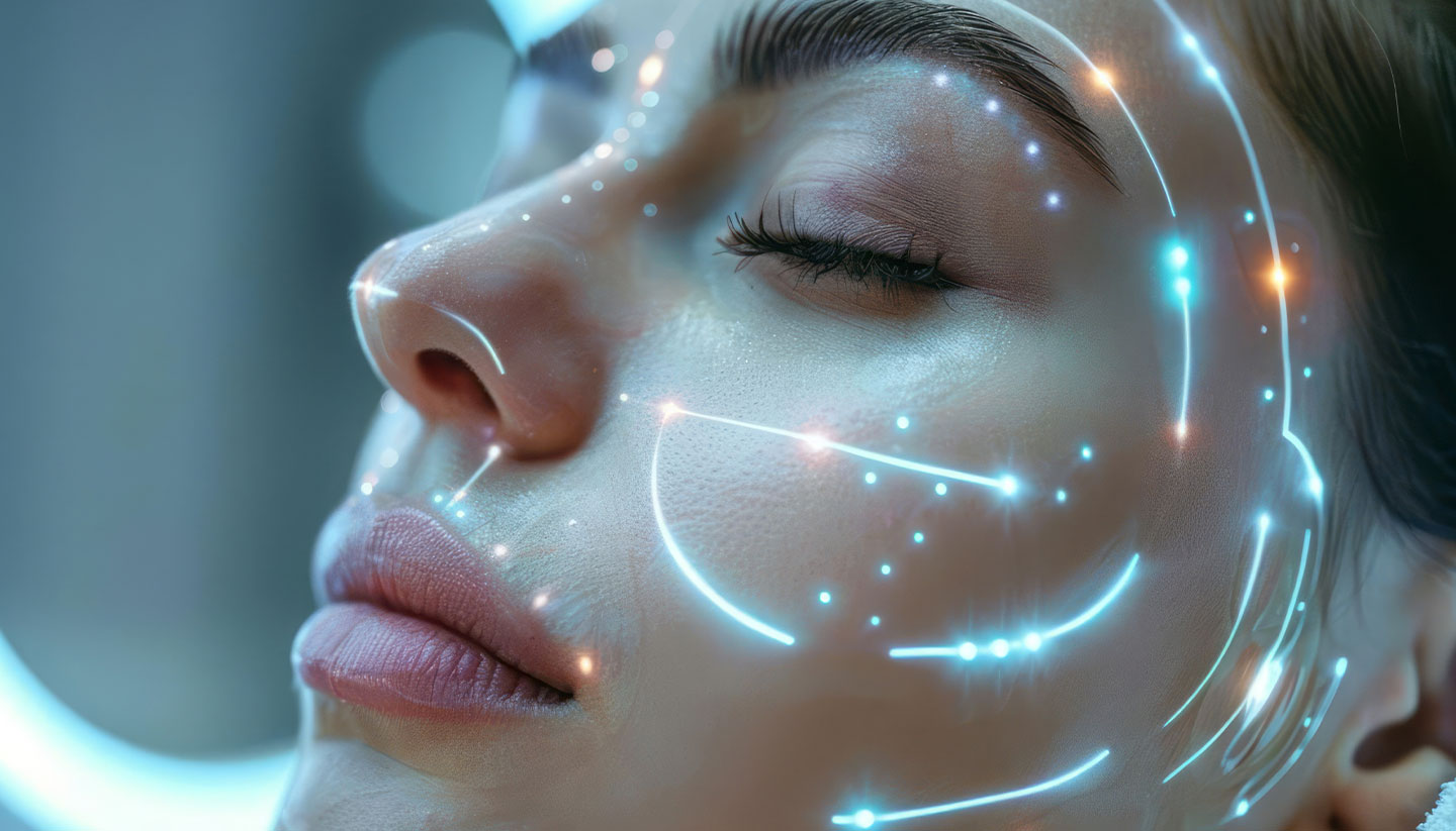
The skin barrier plays a crucial role in protecting the body from harmful environmental factors and maintaining internal hydration. This article explores the structure and functions of the skin barrier, emphasising its interaction with cosmetic products. Methods for analysing barrier health, including transepidermal water loss (TEWL) measurement, Line-Field Confocal Optical Coherence Tomography (LC-OCT) and Raman spectroscopy, are discussed alongside their implications for evaluating product tolerability and efficacy. Additionally, the importance of the hydrolipid film and microbiome in maintaining barrier integrity is highlighted. The article concludes with insights into the development of effective testing protocols and strategies for verifying product efficacy in terms of skin barrier protection or repair.
Introduction
??????????????????
“
“A study in healthy women providing probiotic yogurt for four weeks showed an improvement in emotional responses as measured by brain scans”
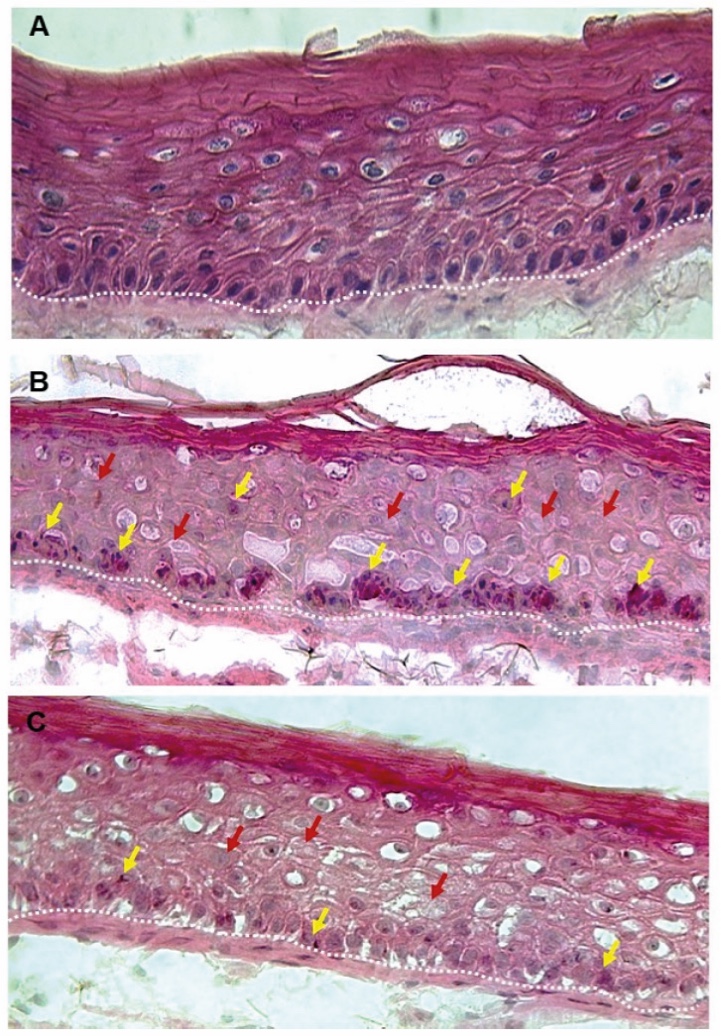
Figure 1. Skin Section with Microbiome. Most microorganisms live in the superficial layers of the stratum corneum and in the upper parts of the hair follicles. Some reside in the deeper areas of the hair follicles and are beyond the reach of ordinary disinfection procedures. There bacteria are a reservoir for recolonization after the surface bacteria are removed.
Materials and methods
Studies of major depressive disorder have been correlated with reduced Lactobacillus and Bifidobacteria and symptom severity has been correlated to changes in Firmicutes, Actinobacteria, and Bacteriodes. Gut microbiota that contain more butyrate producers have been correlated with improved quality of life (1).
A study in healthy women providing probiotic yogurt for four weeks showed an improvement in emotional responses as measured by brain scans (2). A subsequent study by Mohammadi et al. (3) investigated the impacts of probiotic yogurt and probiotic capsules over 6 weeks and found a significant improvement in depression-anxiety-stress scores in subjects taking the specific strains of probiotics contained in the yogurt or capsules. Other studies with probiotics have indicated improvements in depression scores, anxiety, postpartum depression and mood rating in an elderly population (4-7).
Other studies have indicated a benefit of probiotic supplementation in alleviating symptoms of stress. In particular, researchers have looked at stress in students as they prepared for exams, while also evaluating other health indicators such as flu and cold symptoms (1). In healthy people, there is an indication that probiotic supplementation may help to maintain memory function under conditions of acute stress.
The skin is the largest and one of the most important organs of the human body, as it protects the body from external influences such as the penetration of harmful microorganisms, pollution and radiation, as well as preventing excessive loss of water through the skin (1,2). As the primary line of defence, its health directly influences overall well-being. With the increasing demand for high-performance cosmetic products, understanding and measuring the skin barrier's integrity has become vital. This article delves into the structure of the skin barrier, explores advanced methodologies for its analysis, and examines how these insights translate into developing tolerable and effective cosmetic formulations.
Skin barrier structure
The functionality of the skin barrier is centred on its outermost layer, the stratum corneum (SC). Often described as having a ‘bricks and mortar’ structure, this consists of dead keratinised keratinocytes, known as corneocytes, which serve as the ‘bricks’, while the surrounding lipids act as the ‘mortar’.
The ‘bricks’ are better characterised as keratin sponges infused with a natural moisturising factor (NMF), encased within a continuous, highly ordered lamellar lipid phase, largely orthorhombic in structure. Corneocytes are tightly linked by molecular structures called corneo-desmosomes. pH-dependent enzymes are active on the surface of the SC, breaking down these bonds to allow the shedding of corneocytes (3). The SC is also coated in a hydrolipid film, mainly formed by secretions of the sweat and sebaceous glands and thereby consisting of a mixture of water, electrolytes, free fatty acids and lipids from the sebum as well as bactericidal peptides. The hydrolipid film plays a crucial role in various functions, such as preventing TEWL and solute depletion, as well as acting as a defence against pathogens.
Beneath the SC are several epidermal layers where keratinocytes morphologically change and differentiate as they migrate from the stratum basale to the SC. These keratinocytes are interconnected by desmosomes, ensuring cohesion during this process. The epidermis continuously renews itself through desquamation, which is a balancing act between shedding the ‘old’ corneocytes on the surface and keratinocyte proliferation in the stratum basale. This process is important for maintaining the skin’s periodic renewal, and any disturbance can result in a damaged skin barrier (4,5).
Function and dysfunction
The skin barrier acts as the interface between our bodies and the environment, fulfilling a complex and vital protective role. The epidermis is particularly important in this context, offering protection against mechanical, biological, chemical and physical external influences, as well as preventing water loss from the body. This is especially significant given that the human body – which varies in water content depending on age – is composed of approximately 55–75% water (6), in contrast to the relative dryness of the air.
A healthy skin barrier offers protection through four levels of defence: the lipid envelope prevents uncontrolled water loss and solute depletion; the stratum corneum, consisting of corneocytes held together by lipids, provides physical protection against pathogens and mechanical injury; and the chemical/biochemical barrier is formed by lipids, hydrolytic enzymes, acids and antimicrobial peptides. In addition, there is the immunological barrier composed of cellular and humoral constituents, though it will not be discussed in detail here (5).
The hydrolipid film is central to these protective functions. Not only does it keep the skin supple and elastic, aiding mechanical defence, but it also provides protection through the natural microorganisms that inhabit the skin, the microbiome. These organisms have many positive functions for the skin. In addition to producing antimicrobial substances to prevent infection by pathogens, some also have immune system activating properties (7, 8, 9). This biological defence is further supported by the low pH of the skin, which is maintained by compounds such as lactate.
Harmful environmental factors that we are exposed to daily – such as detergents, cleaning products, surfactants, allergen proteases, emulsifiers, diesel exhaust, tobacco, particulate matter, nanoparticles, ozone and microplastics – can degrade the skin barrier by damaging intracellular barrier proteins at intercellular junctions (10). The communication pathway and, consequently, the beneficial relationship between the skin and its microbiome are disrupted when the barrier components of the skin are lost (11, 12). A damaged skin barrier or a disruption to the homeostasis of the skin's microbiome can have various consequences. These include infections, irritations and the emergence of other skin conditions like atopic or contact dermatitis (10, 13, 14).
The skin barrier not only protects but also influences overall well-being through its appearance, texture and even scent. Therefore, it is crucial for the cosmetics industry, among others, to develop products that preserve and even strengthen the microbiome while enhancing the skin barrier’s functionality, as a healthy microbiome and a robust skin barrier are essential for maintaining overall health.
When the skin barrier is damaged, it becomes susceptible to pathogenic, chemical and photochemical agents, resulting in inflammatory reactions that increase skin sensitivity and water loss. This loss of water can lead to skin dryness, both a symptom and a consequence of barrier damage.
Analysing the skin barrier
Consumer demands around cosmetic products are constantly evolving. There is now an increasing focus on products that deliver a range of benefits beyond moisturising, such as protection, nourishment and smoothing. To ensure they are effective and yet do not compromise the skin barrier, these products must undergo comprehensive testing to verify their efficacy and confirm they will not compromise the skin barrier.
Multiple non-invasive methods are now available to scientists looking to assess the tolerability and efficacy of dermatological products. These include:
1. Transepidermal water loss (TEWL) – this technique measures the amount of water released through the skin and serves as the standard non-invasive method for assessing skin barrier integrity. Results are expressed in terms of water mass per unit area and time. Elevated TEWL values indicate areas with a compromised skin barrier. However, it should also be noted that even ‘normal’ skin can exhibit considerable variation in TEWL, so results should be interpreted relatively.
TEWL can be measured using either open or closed chamber systems. In the open chamber system (Figure 1), water vapour from the skin rises vertically through the measuring probe, passing through two or more sensors before exiting the probe. This method provides highly accurate measurements, but it is also extremely sensitive to environmental factors, requiring precise horizontal positioning of the probe. Alternatively, the closed chamber system is more robust and particularly suited for measuring areas of the body that are difficult to position horizontally or where significant airflow is present.
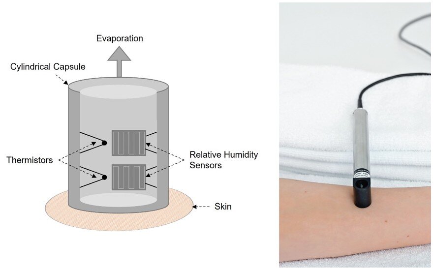
Figure 1. Structure of a sonde for TEWL measurement (modified after Grove et al. (15)) and performance of a TEWL measurement. Transepidermal water loss (TEWL) measurement informs about the epidermal permeability barrier. Low values indicate intact/improved skin function, high values indicate skin barrier abnormalities.
2. Lipbarvis® technology – this technique, previously described by Dähnhardt-Pfeiffer et al., offers detailed insights in the skin barrier’s condition by assessing the SC lipid lamellae structure and the lipid content (16). It delivers both valuable scientific data and marketing-relevant images. A special adhesive is applied to a carrier system and allowed to briefly harden on the skin to remove corneocytes from the surface. Afterwards, the SC’s intracellular lipid lamellae organisation in the samples can be analysed by transmission electron microscopy (Figure 2). A damaged skin barrier is indicated by disrupted or absent lipid lamellar structures.
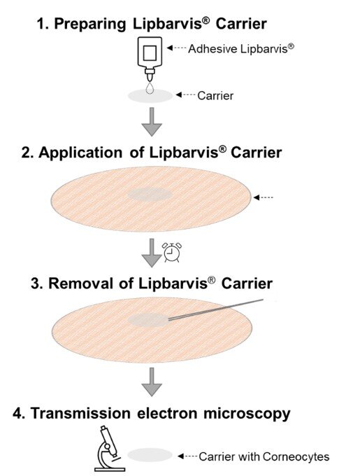
Figure 2. Implementation of the Lipbarvis® technique. Lipbarvis® technology serves the Lipid Barrier Visualisation (LBV).
Conclusion: Upholding Integrity in a Transformative Industry
3. Line-field confocal optical coherence tomography (LC-OCT) – combines the principles of optical coherence tomography (OCT) and confocal reflection microscopy, enabling measurement of SC thickness and providing a unique approach to skin barrier analysis.
Skin is illuminated with a focused line of light, enabling in vivo, cell-resolving imaging. This allows real-time observation of the skin barrier and generates horizontal and vertical slices, as well as three-dimensional images, making it possible to visualise skin structures at the cellular level (17).
By visualising the skin layers in detail, structural changes in skin texture and thickness can be identified, which play a role in evaluating the effectiveness of skincare products but can also be used to diagnose skin diseases such as psoriasis and atopic dermatitis. The formation of new skin tissue and thus the regeneration of the skin barrier after damage can be analysed as well.
4. Raman® spectroscopy – can be used to evaluate the penetration of active ingredients into the skin or to measure the skin’s water gradient. It works by recording the inelastic scattering of light on molecules or solids (18). Skin barrier health is assessed by comparing results before and after treatment. For instance, higher penetration rates of substances like caffeine or a less steep water gradient often indicate a weakened skin barrier.
Each method has its specific strengths, weaknesses and applications, depending on which aspects of the skin barrier are to be examined. The Lipbarvis® technique, for example, is less susceptible to interference from environmental factors such as temperature and humidity, as well as individual factors (e.g., stress), compared to TEWL measurements. While TEWL is an important measurement parameter, providing insights into the structure of the skin barrier, it is also more susceptible to interference and only indirectly measures the barrier function of the skin.
While TEWL measurements provide fast and reproducible results, the Lipbarvis® technology offers clear morphological results that can be correlated with the determination of skin lipids, skin hydration and epidermal proteins. However, analysing the samples can be quite time consuming.
Raman spectroscopy can also be used to investigate skin components and changes, providing detailed molecular information and allowing the analysis of a large number of samples, making it faster to evaluate than data from Lipbarvis® technology. However, analysis can be complicated by fluorescent samples. Furthermore, the devices are expensive and require specialised knowledge to operate and interpret the data.
Analysing the hydrolipid film
The hydrolipid film of the skin can be analysed through its microbiome and molecular components (i.e., lipids, electrolytes, sebum), both of which are crucial for understanding skin health and the impact of external factors like cleansing and skincare products. The microbiome supports skin health by preventing pathogenic colonisation, aiding physiological processes (e.g., keratinocyte differentiation via aryl hydrocarbon receptor (AHR) activation), and therefore maintaining the skin barrier (19). It can be studied using methods such as skin swabs and adhesive films to assess microbial diversity and composition. Since significant changes in the microbiome can harm the skin barrier, evaluating the microbiome-friendliness of products is essential. The microbiome, or alpha diversity, is strongly influenced by skin physiology. When the skin is healthy, moisturised, and the barrier is intact, alpha diversity also increases (19).
The molecular components of the hydrolipid layer, including natural moisturising factors (NMF), can be analysed via aqueous or alcoholic extraction and techniques like Raman spectroscopy. These analyses help reveal the skin barrier's condition by detecting specific substances. However, factors like cleansing and clothing friction can significantly alter these components, highlighting the importance of evaluating products for their effects on the hydrolipid film.
The role of advanced technology in tolerance assessment
A common question within the cosmetics industry is whether we truly need to use these advanced technologies when effective and long-standing methods for tolerance assessment already exist. The answer is yes. While patch tests, in-use studies, and in vitro testing solutions such as red blood cell test and HET-CAM will remain vital parts of the cosmetic scientists’ toolkit, there is a need to monitor the maintenance of the skin barrier, especially as skin is becoming more sensitive to environmental influences. We also require methods to exclude irritant factors, particularly vulnerable populations such as babies, small children and pregnant women.
Tolerability risks that conventional assessments may miss can be identified by enhancing methods like patch tests with measures such as TEWL. This approach increases sensitivity to potential skin barrier damage and helps assess the maintenance of barrier integrity. Other methods, such as microbiome analysis, can also validate claims like ‘microbiome-friendly’, ensuring cosmetic treatments support skin health.
Investigating cosmetics’ impact on the skin barrier
A damaged barrier is essential for investigating product efficacy in regenerating skin barrier function. This damage can be observed naturally, for example, during cold weather or through conditions like atopic dermatitis. However, artificial damage is preferred for cosmetic studies to ensure consistency. This allows researchers to manipulate barrier integrity while working with healthy skin.
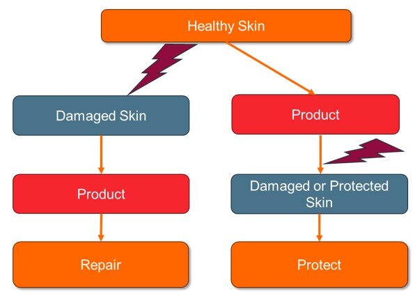
Figure 3. Artificial damage of the skin barrier helps to investigate product efficacy. A noxious agent (shown here as red flashes) is used to damage the skin barrier. The product to be tested either has a regenerative (left) or a protective effect (right) against environmental factors. In the case of the regenerative test design, the skin is damaged by a noxious agent. The test product is then applied in a predetermined regimen and assessed for its ability to repair the skin barrier. Regarding the protective test design, the product is first applied in a predetermined regimen and then artificial damage is produced. It can then be determined whether the product was able to protect the skin barrier or whether the skin barrier was damaged.
Study designs typically focus on regenerative or protective effects, which differ based on the timing of product application. For regenerative studies, the product is applied after damage occurs, while protective studies involve applying the product before damage. The timing is critical in understanding how treatments support barrier recovery or protection (Figure 3).
Damage intensity is another key factor. It can be chronic, occurring over several days, or acute, caused by a single exposure. The severity of the damage depends on the noxious agent used and its exposure time.
The two main methods for artificially damaging the skin barrier are physical and chemical (Table 1). Physical damage includes mechanical methods, and chemical damage is commonly induced using agents that disrupt the skin’s lipid structure, denature proteins, often causing inflammation and cell damage. The severity of chemical damage depends on the specific agent and exposure conditions.
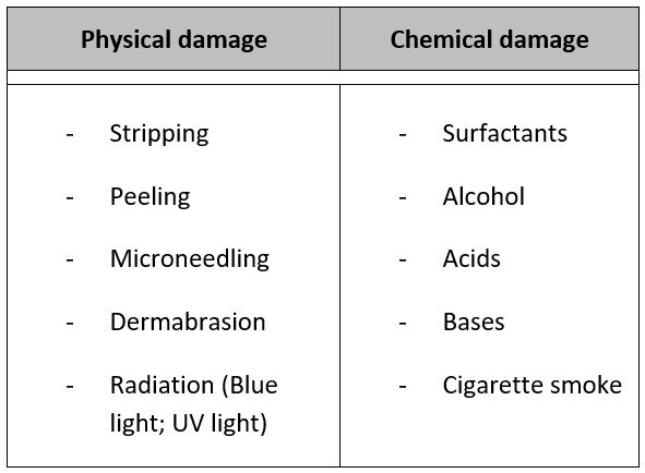
Table 1. Methods for the artificial generation of skin barrier damage. Mechanical methods for physical damage either remove the top layers of the SC without penetrating deep into the epidermis (stripping, peeling) or cause lasting damage to the epidermis (microneedling, dermabrasion). Radiation can also damage all layers of the skin by generating free radicals that trigger inflammation and cell damage. The agents used for chemical damage of the skin can also cause inflammation and cell damage by disrupting the lipid structure and denaturing proteins.
The intensity and duration of damage are critical in studying skin barrier function, helping researchers understand how cosmetic treatments can regenerate or protect the skin.
Choosing the optimal study design
There are various influencing factors to consider when designing a study to assess a product’s efficacy and its impact on the skin barrier. Firstly, we must define the mechanism of action – what the product is designed to do. Secondly, we need to determine the effect we want to demonstrate – repair, protection or maintenance of the skin barrier. These decisions will guide the choice of measurement method and help define the appropriate timeframe in which the product should show its effect. The type and intensity of barrier damage should also align with the product’s mechanism of action.
It is also important to consider the choice of controls: should the product be compared to a placebo or untreated skin? Additionally, it is essential to demonstrate that the selected noxious agent can effectively cause skin damage with the ability to investigate how the damage progresses without product application during the observation period. Finally, the number of test subjects should be considered in relation to the product’s potency.
Creating the optimal study for a new cosmetic product is a complex task. It requires careful consideration of multiple influencing factors to ensure the end product is both safe and effective.
Conclusion
When developing new cosmetic products, it is clear we must not underestimate the importance of the skin barrier. Its resilience and regenerative capabilities are crucial for both health and aesthetics.
As we have seen, our skin is naturally regenerative and protective. Therefore, it is our responsibility to support these functions, ensuring the skin’s barrier remains strong and effective. This requires cosmetic products that not only strengthen and maintain but also repair the skin barrier, addressing its needs to enhance overall well-being.
Equally important is the need for innovative test methods that rigorously validate the microbiome-friendliness, efficacy and safety of new skincare formulations. Protecting and nurturing the skin barrier should be our shared goal, as it is central to preserving its functionality and, by extension, our skin’s health. We therefore must continue to push advancements that help safeguard the skin’s natural defences while improving the products that support it.
Conclusion
The future of cosmetics lies in the continued evolution of holistic approaches which represents a transformative shift in the industry, merging scientific advancements, natural ingredients, and wellness principles. By understanding and embracing the interconnectedness of these elements, the cosmetics industry can cultivate products that not only enhance external beauty but also contribute to the overall well-being of individuals and the planet.
The interplay between beauty from within and topical cosmetics is the key for future products. The integration of biotechnology and green chemistry is revolutionizing cosmetic formulations, offering sustainable and biocompatible alternatives.
Developers can implement blockchain to trace the journey of ingredients from source to product. Nevertheless, the efficacy of the natural products should be scientifically proven. Marketers can communicate transparency as a brand value, and parallelly educate consumers by highlighting how specific ingredients contribute to radiant and healthy skin.
By embracing the synergy between these approaches and leveraging scientific advancements, the cosmetics industry can provide consumers with comprehensive beauty solutions that cater to both internal and external dimensions of beauty.
Surfactant Applications
The application area lends itself particularly well to the use of AI. Active today in this area is the US company Potion AI (6). The company provides AI-powered formulation tools for beauty and personal care R&D. Their offerings include Potion GPT, next generation ingredient and formula databases and AI document processing. Potion’s work could have a significant impact on the entire surfactant value chain, from raw material suppliers to end consumers. By using their GPT technology, they can help target work toward novel surfactant molecules that have optimal properties for specific applications. By using their ingredient and formula databases, they can access and analyze a vast amount of data on surfactant performance, safety, and sustainability. By using their AI document processing, they can extract and organize relevant information from patents, scientific papers, and regulatory documents. These capabilities could enable Potion AI's customers to design and optimize surfactant formulations that are more effective, eco-friendly, and cost-efficient. A particularly interesting application for this type of capability is deformulation.
Deformulation is the process of reverse engineering a product's formulation by identifying and quantifying its ingredients. Deformulation can be used for various purposes, such as quality control, competitive analysis, patent infringement, or product improvement. However, deformulation can be challenging, time-consuming, and costly, as it requires sophisticated analytical techniques, expert knowledge, and access to large databases of ingredients and formulas.
AI can potentially enhance and simplify the deformulation process by using data-driven methods to infer the composition and structure of a product from its properties and performance. For example, AI can use machine learning to learn the relationships between ingredients and their effects on the product's characteristics, such as color, texture, fragrance, stability, or efficacy. AI can also use natural language processing to extract and analyze information from various sources, such as labels, patents, literature, or online reviews, to identify the possible ingredients and their concentrations in a product.
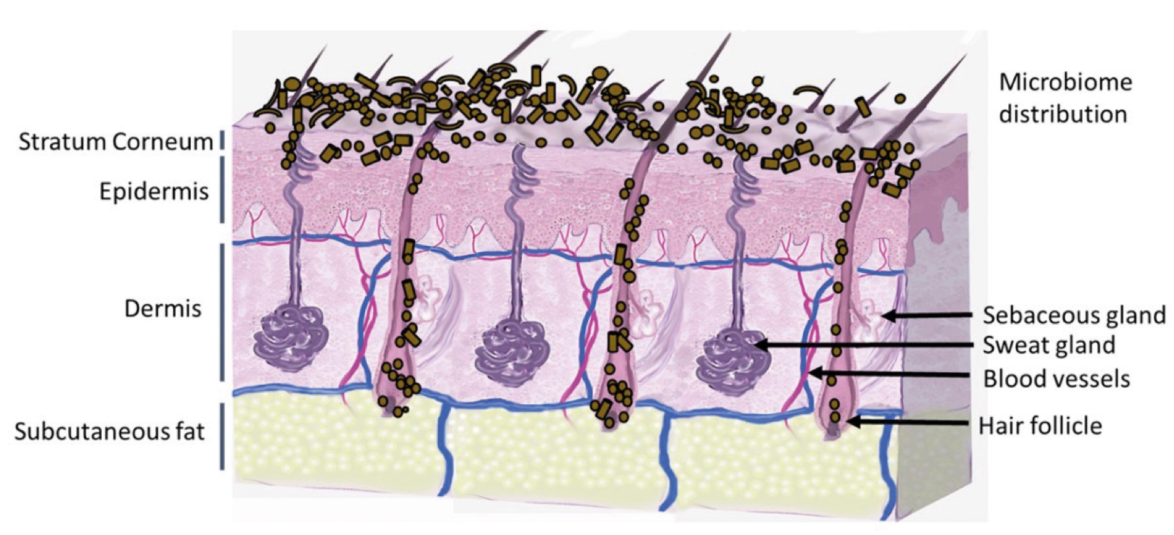
Figure 2. Skin Section with Microbiome. Most microorganisms live in the superficial layers of the stratum corneum and in the upper parts of the hair follicles. Some reside in the deeper areas of the hair follicles and are beyond the reach of ordinary disinfection procedures. There bacteria are a reservoir for recolonization after the surface bacteria are removed.
About the Author
Doerte Segger
 Doerte is Laboratory and Customer Service Manager for dermatological testing at SGS INSTITUT FRESENIUS GmbH. With 30 years of professional experience, including her role as Technical Director of a CRO and her long-standing involvement in various expert committees within the cosmetics industry, she is a recognised expert in developing optimised study designs for tolerability and efficacy studies using both classic and innovative testing methods. Doerte holds a degree in Biochemistry and completed her Dr. rer. nat. in the field of Cosmetic Sciences.
Doerte Segger
Lab and Customer Service Manager, Health & Nutrition, SGS INSTITUT FRESENIUS GmbH, Hamburg, Germany

References and notes
- Proksch E, Brandner J M, & Jensen J M. The skin: an indispensable barrier. Experimental dermatology. 2008;17(12); 1063-1072.
- Lefèvre-Utile A, Braun C, Haftek M, Aubin F. Five functional aspects of the epidermal barrier. International journal of molecular sciences. 2021;22(21); 11676.
- Rawlings A V. Recent advances in skin ‘barrier’ research. Journal of Pharmacy and Pharmacology. 2010; 62(6); 671-677.
- Hänel K H, Cornelissen C, Lüscher B, Baron JM. Cytokines and the Skin Barrier. International Journal of Molecular Sciences. 2013; 14(4); 6720-6745.
- Baroni A, Buommino E, De Gregorio V, Ruocco E, Ruocco V, Wolf R. Structure and function of the epidermis related to barrier properties. Clinics in Dermatology. 2012; 30(3); 257-262.
- Popkin B M, D'Anci K E, Rosenberg I H. Water, hydration, and health. Nutrition reviews. 2010; 68(8); 439-458.
- Karnwal A, Shrivastava S, Al-Tawaha A R M S, Kumar G, Singh R, Kumar A, et al. Microbial biosurfactant as an alternate to chemical surfactants for application in cosmetics industries in personal and skin care products: a critical review. BioMed Research International. 2023; 2023(1); 2375223.
- Ahle C M, Stødkilde K, Poehlein A, Bömeke M, Streit W R, Wenck H, et al. Interference and co-existence of staphylococci and Cutibacterium acnes within the healthy human skin microbiome. Communications biology. 2022; 5(1); 923.
- Yang Y, Qu L, Mijakovic I, Wei Y. Advances in the human skin microbiota and its roles in cutaneous diseases. Microbial Cell Factories. 2022; 21(1); 176.
- Strugar T L, Kuo A, Seité S, Lin M, Lio P. Connecting the dots: from skin barrier dysfunction to allergic sensitization, and the role of moisturizers in repairing the skin barrier. Journal of drugs in dermatology. 2019; 18(6), 581-581.
- Baurecht H, Rühlemann M C, Rodríguez E, Thielking F, Harder I, Erkens A S, et al. Epidermal lipid composition, barrier integrity, and eczematous inflammation are associated with skin microbiome configuration. Journal of Allergy and Clinical Immunology. 2018; 141(5); 1668-1676.
- Uberoi A, Bartow-McKenney C, Zheng Q, Flowers L, Campbell A, et al. Commensal microbiota regulates skin barrier function and repair via signaling through the aryl hydrocarbon receptor. Cell host & microbe. 2021; 29(8); 1235-1248.
- Sözener Z C, Cevhertas L, Nadeau K, Akdis M, Akdis C A. Environmental factors in epithelial barrier dysfunction. Journal of allergy and clinical immunology. 2020; 145(6); 1517-1528.
- Schwartz J, Friedman A J. Exogenous Factors in Skin Barrier Repair. Journal of drugs in dermatology. 2016; 15(11); 1289-1294.
- Poster: Grove G, Zerweck, Damia & C. A Practical Guide to Computerized Evaporimetry (http://www.cyberderm-inc.com/uploads/6/3/5/7/63578583/a_practical_guide_to_computerized_evaporimetry.pdf; 2023, May 25)
- Daenhardt-Pfeiffer S, Surber C, Wilhelm K P, Daenhardt d, Springmann G, Boettchen M, Foelster-Holst R. Noninvasive stratum corneum sampling and electron microscopical examination of skin barrier integrity: Pilot study with a topical glycerin formulation for atopic dermatitis. Skin pharmacology and physiology. 2012; 25(3); 155-161
- Latriglia F, Ogien J, Tavernier C, Fischman S, Suppa M, Perrot J L, Dubois A. Line-field confocal optical coherence tomography (LC-OCT) for skin imaging in dermatology. Life. 2023; 13(12); 2268.
- Lademann J, Meinke M C, Patzelt A, Darvin M E. Raman-Spektroskopie in der Dermatologie. Nichtinvasive physikalische Diagnostik in der Dermatologie. 2016; 103-115.
- Lee HJ, Kimm M. Skin Barrier Function and the Microbiome. International Journal of Molecular Sciences. 2022; 23(21); 13071.