Peptides
Skin care
KEYWORDS
Peptides;
Cell culture;
Paeonia suffruticosa;
Retinol-like;
ECM
peer-reviewed
Peptides from Paeonia suffruticosa cell cultures as a natural Alternative to Retinoids with ECM-Boosting and Barrier-Preserving Benefits
Annalisa Tito1*, Danila Falanga1, Chiara Niespolo1, Ritamaria Di Lorenzo2, Sonia Laneri2 and Maria Gabriella Colucci1-3
*Corresponding author
- Arterra Bioscience SpA, Naples, Italy
- Department of Pharmacy, University of Naples Federico II, Naples, Italy
- Vitalab Srl, Naples, Italy
ABSTRACT: Retinol is one of the most effective and widely used anti-aging ingredients; however, its harshness often causes irritation, particularly in sensitive skin. Consequently, the search for plant-derived retinol alternatives with comparable efficacy but improved tolerability is ongoing. This study investigates a peptidic extract, obtained with a patented procedure, from cell walls of Paeonia suffruticosa cell cultures that activates retinoic acid receptor boosting extracellular matrix proteins under basal and stressed conditions. Unlike retinoic acid, the Paeonia extract promoted the expression of tight junction proteins such as claudin-1, highlighting its favorable profile for maintaining skin barrier function. A clinical study comparing a formulation containing this extract with one containing 0.3% retinol revealed the superior efficacy of the Paeonia suffruticosa peptidic extract.
Introduction
Retinol undoubtedly represents the benchmark ingredient in antiaging skin care for its ability to boost collagen production and repair photo-induced skin damages. In the skin, retinol is metabolically converted into retinoic acid, its biologically active form which exerts its effects by binding the Retinoic Acid Receptor (RAR) and activating downstream signal cascades involved in cell differentiation, proliferation and extracellular matrix (ECM) remodeling (1). During UV exposure, retinoic acid suppresses the activation of AP-1 transcription factors, inhibiting the expression of matrix metalloproteinases (MMPs) responsible for collagen and elastin degradation, while simultaneously promoting the synthesis of structural ECM proteins (2). However, despite its efficacy, retinol is often associated with a considerable risk of irritation, dryness, and erythema particularly in individuals with sensitive or compromised skin (3). These side effects have driven scientific and industrial interest to the identification of alternative and natural molecules able to mimic the biological activity of retinol without its side effects. Among retinol-like ingredients, peptides have gaining attention due to their ability to promote collagen and glycosaminoglycan production in skin cells (4). Moreover, our group described the application of peptide and sugar mixtures generated by partial digestion of plant tissue culture cell wall glycoproteins in cosmetic formulations, showing beneficial effects on skin cells and on human skin in vivo (5, 6, 7, 8). In this study we reported for the first time the involvement of retinoic acid pathway in the biological mechanisms induced by a peptide sugar mixture derived from Paeonia suffruticosa liquid suspension culture. We demonstrated that Paeonia peptides extract activates the Retinoic Acid Receptor and, as consequence, protects and repairs collagen and fibrillin fibers from damages induced by photo-aging as shown by in vitro and ex vivo test, respectively. Clinical tests demonstrated that Paeonia peptides improved skin texture and increased skin firmness after 14 and 28 days of treatment, outperforming retinol used as the benchmark.
Materials and Methods
Paeonia suffruticosa cell culture extract (Paeonia Peptides). An extract rich in peptides and sugars was obtained by Paeonia suffruticosa cell cultures according to the protocol described by Apone et al (8).
Cell line and skin explants. HaCaT (Human keratinocytes) and HDF (Human Dermal Fibroblast) were maintained in Dulbecco’s modified Eagle’s medium (DMEM) plus 10% FBS at 37°C under 5% CO2. Skin explants were obtained from healthy female donors (40, 49 and 50 years old) following breast surgery. All donors had given their written informed consent for the use of the skin tissues (according to the Helsinki Declaration). Samples were cut with punch biopsy curettes (8mm) and cultured in 24-transwell plates in DMEM plus FBS plus antibiotics at 37°C in 5% CO2. Ex-vivo tests were performed in three biological and technical replicates: three punches derived from three different donors.
In vitro assays.
Activation of RAR
HaCaT cells were transfected with a reporter-based vector encoding for Retinoic Acid Receptor Element (RARE). 24 hours after transfection the cells were treated with Paeonia peptides or Retinoic acid and after an additional 24 hours, were lysed and processed using Steady-Glo® Luciferase assay system (Promega Corporation). Luminescence associated with receptor activation was measured using the Victor Nivo multimode plate reader (Perkin Elmer).
Quantification of collagen by ELISA after IR-induced damage
HDF were treated for 16 hours with Paeonia peptides and positive control and subsequently irradiated with Infrared radiation (88 J/cm2). Following irradiation cells were treated again with the samples and incubated for additional 72 hours. At the end of the incubation period, cells were processed for ELISA to quantify the content of mature type I collagen by using Monoclonal Anti-Collagen Type I (C2456, Sigma Aldrich). Collagen I levels were determined by measuring absorbance with a Victor Nivo plate reader (Perkin Elmer).
Ex vivo assays
ImmunoHistoFluorescence for fibrillin-1 after UVA-induced damage
Photoaging was induced by sequential UVA irradiation of skin explants at 22 J/cm², applied once daily for 3 consecutive days. Following irradiation, skin samples were treated for an additional 5 days with Paeonia peptides or retinoic acid for comparison. At the end of the treatment period, skin punches were fixed in paraformaldehyde and cryopreserved. Frozen sections (5 μm) were prepared using a CM1520 cryostat (Leica Microsystems), rinsed with PBS, and incubated with the appropriate fibrillin-1 primary antibody (MA512770, Thermo Fisher Scientific), followed by nuclear staining with DAPI (1 μg/mL). Images were acquired using a fluorescence microscope and analyzed with ImageJ software. Fluorescence intensity associated with candelabra-like structures, characteristic of fibrillin fibers, was measured.
ImmunoHistoFluorescence for claudin-1
For claudin-1 quantification, ex vivo skin explants were treated with Paeonia peptides or with retinoic acid for 24 hours and then processed for immunohistofluorescence (as described above) using claudin-1 Polyclonal Antibody (51-9000, Thermo Fisher Scientific).
Statistical analysis
All data represent the mean ± standard deviation (SD) of at least three independent experiments. Statistical significance was determined using t-test and the number of asterisks in the graphs indicate the level of statistical significance (*** p-value < 0.001; ** 0.001 < p < 0.01; * 0.01 < p < 0.05). The values are considered significant when the p-value is ≤ 0.05.
Clinical study
To further investigate the biological actions, a randomized, double blind clinical study was conducted on 60 female volunteers (Fitzpatrick skin types I-III) aged between 25 and 55 years. All participants exhibited facial skin concerns, including pigmentation irregularities, fine lines, wrinkles, and uneven skin texture.
The volunteers were divided into three groups, one treated with an emulsion containing Paeonia peptides, one with a formula containing 0,3% of retinol and another with a placebo. Over a 28-day period, each group applied one of the assigned formulations to the face twice daily.
Treatment efficacy was assessed through the evaluation of the skin texture and skin firmness after 14 and 28 days by product application (D14, and D28).
In particular, skin texture was assessed using the VISIA® 7th system (Canfield Scientific Inc., USA), an imaging device that enables the objective detection and quantification of skin surface irregularities, including roughness and microrelief alterations. Whereas skin firmness was evaluated using the Cutometer® MPA 580 (Courage + Khazaka Electronic GmbH, Germany).
The tests adhered to the principles of the Helsinki Declaration and the Colipa Guidelines.
Statistical analysis in Clinical trial
A sample size of 60 panelists, randomly assigned (approximately 20 subjects per group), was considered sufficient to achieve adequate statistical power for detecting differences between the two treated groups and placebo.
Inter-group differences were assessed using the ANOVA test, while intra-group differences, expressed as average percentage variations compared to baseline, were analyzed using the Student t-test. The significance level for all analyses was set at p-value < 0.05.
Results
During the screening for Retinoic acid receptor (RAR) modulators we selected a peptidic extract derived from a cell wall preparation of Paeonia suffruticosa cell culture. The peptides and sugars mixture was obtained by a cell wall extraction from liquid suspension cultures of P. suffruticosa cells, following a patented protocol (WO2007104489A1) already described by Apone et al. (8). Figure 1 reports the results of a cell-based RARE reporter assay. As shown, the extract activates RAR in a dose-dependent manner, with receptor activity reaching 20% at the lower dose and 35% at the higher dose.
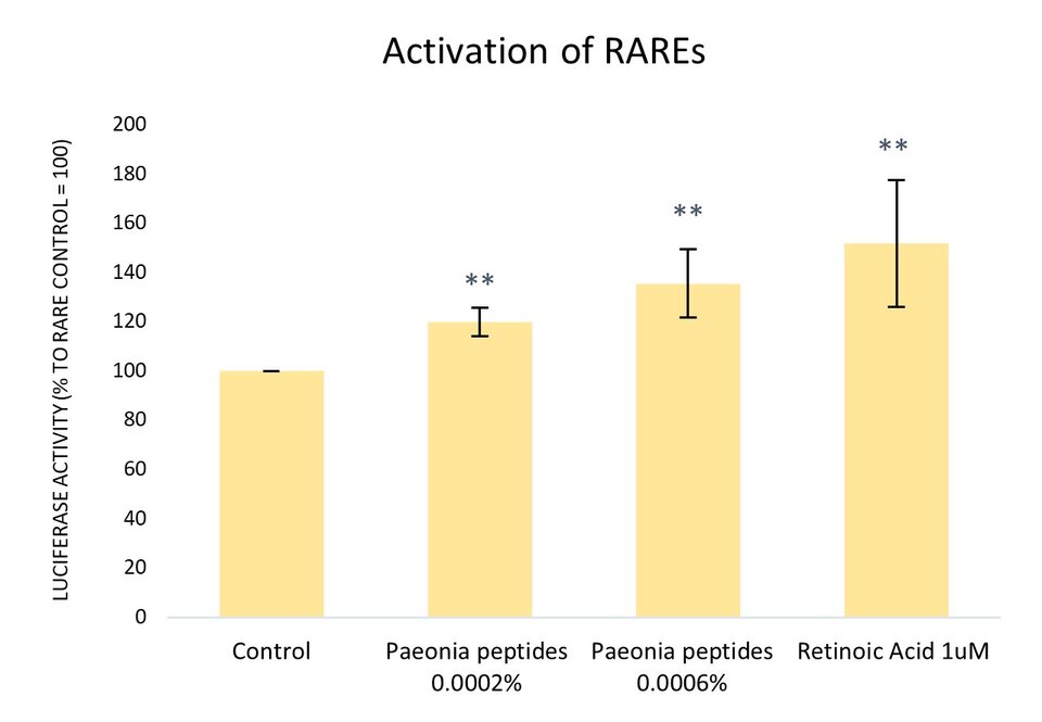
Figure 1. Activation of Retinoic Acid Responsive Elements (RAREs) measured through luciferase activity.
Graph showing the ability of P. suffruticosa peptides to activate RARE in transfected HaCaT cells. The bars represent the mean ± standard deviations, and the asterisks indicate significant variations according to Student’s t-test (* p < 0.05, ** p < 0.01, *** p < 0.001).
Considering the activation of Retinoic acid Receptor, next we evaluated the effect of P. suffruticosa peptides on collagen photoprotection. Human dermal fibroblasts, treated with Paeonia peptides or with retinoic acid, were irradiated with InfraRed to damage collagen proteins and then treated with the samples for further 72 hours.
As shown in Figure 2, the content of mature collagen in InfraRed irradiated fibroblasts decreased by 70% compared to untreated cells but in the presence of Paeonia peptides, collagen percentage is rescued to the basal level. This effect is also more evident than that generated by the retinoic acid.
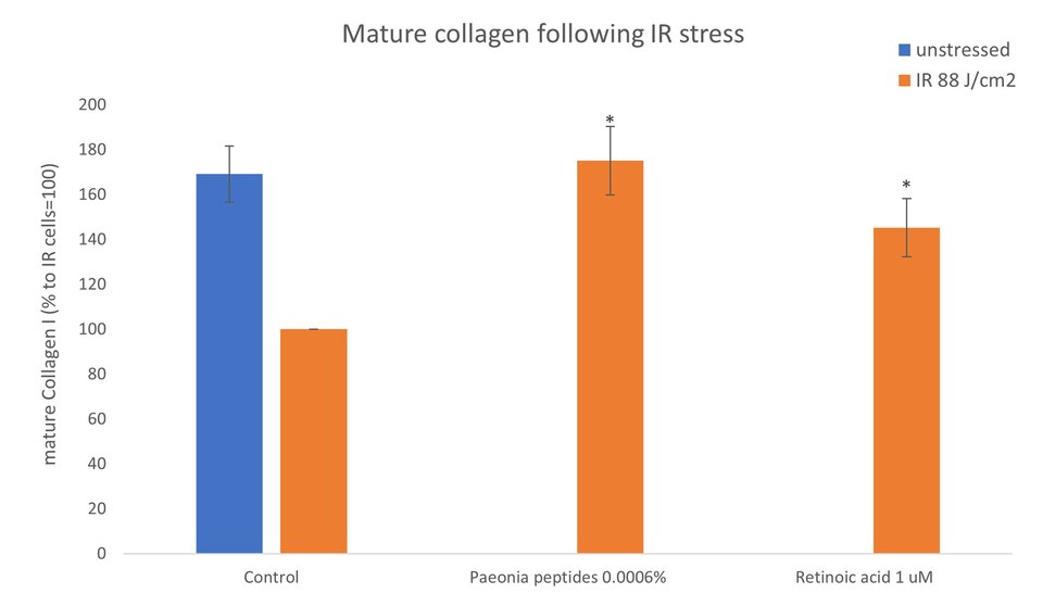
Figure 2. Quantification of mature collagen in HDF irradiated with infrared.
Graph showing the ability of Paeonia peptides to maintain the level of mature collagen up to basal level (non-irradiated cells). The bars represent the mean ±standard deviations, and the asterisks indicate significant variations according to Student’s t-test (* p < 0.05, ** p < 0.01, *** p < 0.001).
The positive effect on photoaging was confirmed in an ex vivo test monitoring the quantity and the correct structure of fibrillin-1, one of the ECM proteins involved in skin elasticity and used as a marker of photoaging. As shown in the Figure 3 the quantity of fibrillin-1 decreased by 56% in irradiating skin explants reflecting the typical degradation of extracellular matrix components associated with photoaging. However, the treatment with Paeonia peptides restored this reduction, repairing fibrillin-1 proteins at levels comparable to non-irradiated controls. This repairing effect was found to be equivalent to that observed with retinoic acid treatment, highlighting the extract's potential as an effective agent for photoaging issues.
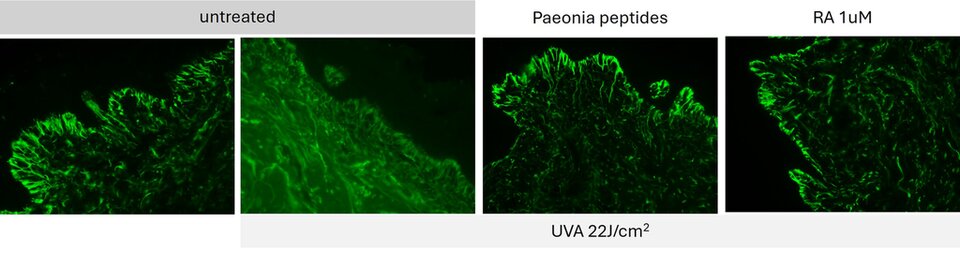
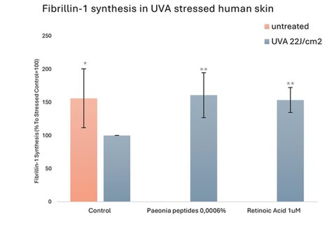
Figure 3. Fibrillin-1 in UVA irradiated human skin explants: a) representative pictures; b) quantification of fluorescence.
Detection of fibrillin-1 in skin sections. The bars represent the standard deviations, and the asterisks indicate significant variations according to Student’s t-test (* p < 0.05, ** p < 0.01, *** p < 0.001).
Since retinol is commonly associated with skin barrier disruption, we also assessed the effect of our extract on skin barrier integrity focusing on detection of claudin-1, one of proteins that compose tight junctions between keratinocytes. Skin explants treated with Paeonia peptides or with retinoic acid were processed for immunohistofluorescence against claudin-1 and the results were reported in the Figure 4. Notably, unlike retinoic acid, which compromises claudin proteins, Paeonia peptides increased the expression of the tight junction protein claudin-1 in human skin explants. This effect suggests a link to enhanced barrier integrity. Overall, these findings suggest that Paeonia extract may serve as a natural and safer alternative to retinoid-like compounds for promoting ECM production while supporting skin barrier function.
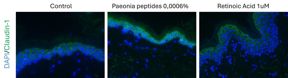
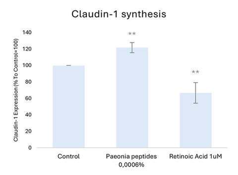
Figure 4. Ex vivo immunofluorescence of epidermal tight-junction claudin-1: a) representative pictures; b) quantification of fluorescenc.
Detection of claudin-1 in skin sections. The bars represent the standard deviations, and the asterisks indicate significant variations according to Student’s t-test (* p < 0.05, ** p < 0.01, *** p < 0.001).
Clinical study
The treatment with the formula containing Paeonia peptides and that containing retinol resulted in a marked improvement in skin smoothness. Paeonia peptides reduced surface irregularities, showing a 18% decrease compared to baseline at D14, and a 15% reduction at D28. The retinol-treated group also showed a 16% improvement in skin texture at D14, followed by a smaller, non-significant 8% reduction at D28.
In contrast, the placebo group did not exhibit any improvement in skin texture. Corresponding percentage changes from baseline were represented in Figure 5.
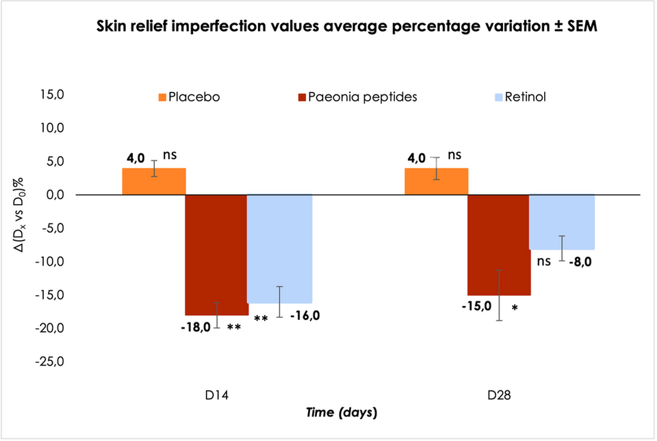
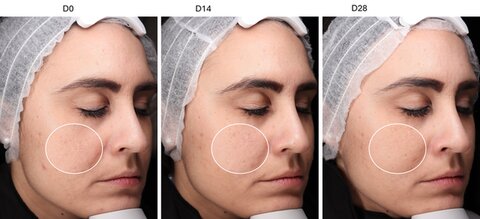
Figure 5. a) Skin relief percentage variation during the treatment with Paeonia peptides, Retinol or placebo, respectively, at each follow-up session (D14 and D28). The baseline data served as the reference value for each analysis. Data were shown as mean ± SEM (n=20). (intra-group difference t-test: ns = not significant, * p < 0.05, and ** p < 0.01 vs D0; inter-group difference ANOVA test: $$$ p < 0.001 vs placebo). b) Representative VISIA 7th images of a 34-year-old subject following treatment with Paeonia peptides.
In addition, both the Paeonia- and retinol-treated groups showed increased skin firmness, reflected by a decrease in the R0 parameter, with lower R0 values corresponding to greater firmness. In particular, Paeonia peptides exhibited a marked firming effect, with a 20% reduction in R0 at D14, which further improved to a 26% decrease after 28 days of treatment (Figure 6). In comparison, the retinol-treated group showed an 18% reduction in R0 at D14 and a 19% reduction at D28. In contrast, the placebo group showed negligible effects on skin firmness, with not significant R0 variation.
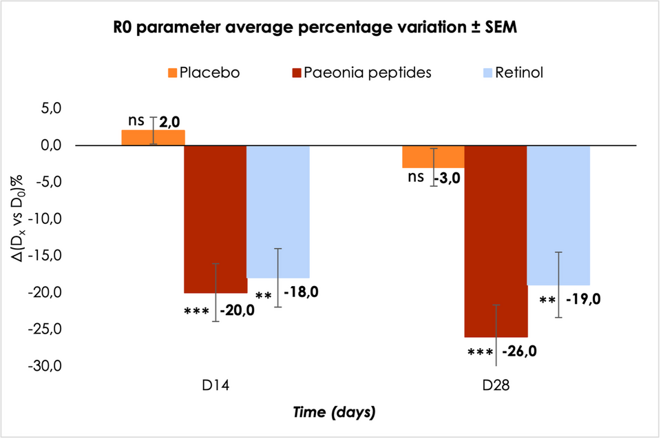
Figure 6. R0 parameter percentage variation during the treatment with Paeonia peptides, Retinol or placebo, respectively, at each follow-up session (D14 and D28). The baseline data served as the reference value for each analysis. Data were shown as mean ± SEM (n=20). (intra-group difference t-test: ns = not significant, ** p < 0.01, and *** p < 0.001 vs D0; inter-group difference ANOVA test: $$ p < 0.01, $$$ p < 0.001 vs placebo).
Discussion
The present study provides the first evidence that a peptides and sugars rich extract derived from Paeonia suffruticosa cell cultures exerts retinol-like biological activity modulating retinoic acid receptor pathway. The functional consequences of RAR activation were confirmed by the marked protective and reparative effects on ECM components under photoaging conditions as demonstrated by in vitro experiments on collagen I and in ex vivo on fibrillin-1 protein. Treatment with Paeonia peptides effectively repaired fibrillin networks, restoring both protein levels and the characteristics candelabra like structures to values comparable to those observed in non-irradiated skin. Notably, the magnitude of this effect was equivalent to that obtained with retinoic acid. A critical aspect distinguishing Paeonia peptides with classical retinoids emerged from the analysis of its effect on one of the most important epidermal barrier markers. Ex vivo experiments revealed that retinoic acid reduces claudin1, impairing tight junctions and epidermal barrier, while Paeonia peptides increased the expression of this marker. This dual action, promotion of ECM repair alongside support of epidermal barrier integrity, represents a major advantage over conventional retinoids and addresses one of their principal limitations in long-term cosmetic use. The clinical outcomes are fully consistent with the biological data. In a randomized, double blind, placebo-controlled study, topical application of a formula containing Paeonia peptides resulted in significant improvements in skin texture and firmness after just 14 days, with effects further enhanced at 28 days. Compared to retinol, Paeonia peptides demonstrated superior performance, improving skin firmness approximately 1,4-fold and smoothing texture up to 1,9-fold more effectively.
Conclusion
Taken together, all the results indicate that Paeonia peptides derived from Paeonia suffruticosa cell cultures act as a novel, plant-based retinol like ingredient. By activating the retinoic acid pathway, protecting and repairing ECM components, and simultaneously reinforcing epidermal barrier function, this extract offers a compelling alternative to classical retinol for next generation cosmetic formulation.
About the Author
Annalisa Tito is the Director of the Molecular and Cell Biology Laboratory at Arterra Bioscience S.p.A. She is responsible of the internal biological efficacy platform, supporting the substantiation of active ingredient claims through innovative methodologies.
She is co-author of more than 40 scientific publications and co-inventor of several patents. Currently, based on her expertise in molecular and cell biology, Annalisa coordinates the development of active ingredients for Vitalab Srl, an innovative joint venture between Arterra Bioscience and Intercos Group.
Annalisa Tito
Arterra Bioscience SpA, Naples, Italy

References and notes
- Gudas LJ. Retinoids metabolism: new insights. Journal of Molecular Endocrinology Available from https://doi.org/10.1530/JME-22-0082
- Schüle, R et al. Retinoic acid is a negative regulator of AP-1-responsive genes. Proceedings of the National Academy of Sciences of the United States of America, 88(14), 6092–6096. Available from https://doi.org/10.1073/pnas.88.14.6092
- Zhong J et al. Topical retinoids: Novel derivatives, nano lipid-based carriers, and combinations to improve chemical instability and skin irritation. Journal of Cosmetic Dermatology. Available from https://doi.org/10.1111/jocd.16415
- Pintea A et al. Peptides: Emerging Candidates for the Prevention and Treatment of Skin Senescence: A Review. Biomolecules 2025. Available from https://doi.org/10.3390/biom15010088
- Falanga D. et al. Effetto migliorativo sull’integrità cutanea di un estratto biotecnologico di Scabiosa arvensis. Available from https://www.ceceditore.com/innovazione-in-botanicals-2-2025-2/
- Tito A et al. Skin rejuvenating properties of a powerful blend of Gardenia jasminoides peptides and flavonoids. HPC Today Available from https://tks-hpc.h5mag.com/hpc_today_1_2024/skin_care_skin_rejuvenating_properties_of_a_powerful_blend_of_gardenia_jasminoides_peptides_and_flavonoids
- Tito A et al. The Growth Differentiation Factor 11 is Involved in Skin Fibroblast Ageing and is Induced by a Preparation of Peptides and Sugars Derived from Plant Cell Cultures. Molecular Biotechnology. Available from 10.1007/s12033-019-00154-w https://link.springer.com/article/10.1007/s12033-019-00154-w?utm_source=researchgate.net&utm_medium=article
- Apone F et al. A mixture of peptides and sugars derived from plant cell walls increases plant defense responses to stress and attenuates ageing-associated molecular changes in cultured skin cells. Journal of Biotechnology. Available from doi:10.1016/j.jbiotec.2009.11.021 https://www.sciencedirect.com/science/article/abs/pii/S0168165609005471?via%3Dihub This work was supported by the EU-H2020 through the project “Innovative high-value cosmetic products from plants and plant cells” (InnCoCells, grant number 101000373).